Subphylum Chelicerata
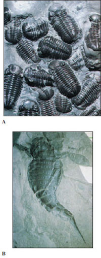 |
| Figure 18-1 Fossils of early arthropods. A, Trilobite fossils, dorsal view. These animals were abundant in mid-Cambrian period. B, Eurypterid fossil. Eurypterids flourished in Europe and North America from Ordovician to Permian periods. |
Chelicerate arthropods are an ancient group that includes eurypterids (extinct), horseshoe crabs, spiders, ticks and mites, scorpions, and sea spiders. They are characterized by having six pairs of appendages that include a pair of chelicerae, a pair of pedipalps, and four pairs of walking legs (a pair of chelicerae and five pairs of walking legs in horseshoe crabs). They have no mandibles and no antennae. Most chelicerates suck liquid food from their prey.
Class Merostomata
Class Merostomata is represented by eurypterids, all now extinct, and xiphosurids, or horseshoe crabs, an ancient group sometimes called “living fossils.”
Subclass Eurypterida
The eurypterids, or giant water scorpions (Figure 18-1B) were the largest of all fossil arthropods, some reaching a length of 3 m. Their fossils occur in rocks from the Ordovician to the Permian periods. They had many resemblances to marine horseshoe crabs (Figure 18-2) and also to scorpions, their land counterparts. Their head had six fused segments and bore both simple and compound eyes and six pairs of appendages. Their abdomen had 12 segments and a spikelike telson.
Ideas regarding their early habitats differ. Some authorities believe eurypterids evolved mainly in fresh water; others hold that they arose in brackish lagoons.
Subclass Xiphosurida: Horseshoe Crabs
Xiphosurids are an ancient marine group that dates from the Cambrian period. Our common horseshoe crab Limulus (L. limus, sidelong, askew) (Figure 18-2) goes back practically unchanged to the Triassic period. Only three genera (five species) survive today: Limulus, which lives in shallow water along the North American Atlantic coast; Carcinoscorpius (Gr. karkinos, crab, + skorpion, scorpion), along the southern shore of Japan; and Tachypleus, (Gr. tachys, swift, + pleutes, sailor), in the East Indies and along the coast of southern Asia. They usually live in shallow water.
Xiphosurids have an unsegmented, horseshoe-shaped carapace (hard dorsal shield) and a broad abdomen, which has a long telson, or tailpiece. Their cephalothorax bears five pairs of walking legs and a pair of chelicerae, whereas their abdomen has six pairs of broad, thin appendages that are fused in the median line (Figure 18-2). On some of the abdominal appendages, book gills (flat, leaflike gills) are exposed. There are two compound and two simple eyes on the carapace. The horseshoe crab swims by means of its abdominal plates and can walk with its walking legs. It feeds at night on worms and small molluscs, which it seizes with its chelicerae.
During the mating season horseshoe crabs come to shore at high tide to mate. A female burrows into sand where she lays eggs, with one or more smaller males following closely to add sperm to the nest before the female covers it with sand. Eggs are warmed by the sun and protected from waves until young larvae hatch and return to the sea by another high tide. Larvae are segmented and are often called “trilobite larvae” because they resemble trilobites, to which xiphosurids may be related.
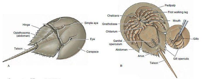 |
| Figure 18-2 A, Dorsal view of horseshoe crab Limulus (class Merostomata). They grow to 0.5 m in length. B, Ventral view. |
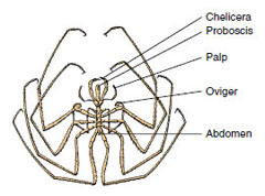 |
| Figure 18-3 Pycnogonid, Nymphon sp. In this genus all anterior appendages (chelicerae, palps, and ovigers) are present in both sexes, although ovigers are often not present in females of other genera. |
Class Pycnogonida: Sea Spiders
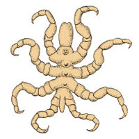
Some sea spiders are only a few millimeters long, but others are much larger. They have small, thin bodies and usually four pairs of long, thin walking legs. In addition, they have a feature unique among arthropods: somites are duplicated in some groups, so that they possess five or six pairs of legs instead of the four pairs normally characteristic of arachnids. Males of many species bear a subsidiary pair of legs (ovigers) (Figure 18-3) on which they carry developing eggs, and ovigers are often absent in females. Many species also are equipped with chelicerae and palps.
Their mouth is at the tip of a long proboscis, which sucks juices from cnidarians and soft-bodied animals. Most pycnogonids have four simple eyes. Their circulatory system is limited to a simple dorsal heart, and excretory and respiratory systems are absent. The long, thin body and legs provide a large surface, in proportion to volume, that is evidently sufficient for diffusion of gases and wastes. Because of the small size of the body, the digestive system sends branches into the legs, as do the gonads.
 |
| Figure 18-4 Pycnogonum, a pycnogonid with relatively short legs. Females of this genus have neither chelicerae nor ovigers and males have ovigers. |
Sea spiders are found in all oceans, but they are most abundant in polar waters. Pycnogonum (Figure 18-4) is a common intertidal genus found on both Atlantic and Pacific coasts of the United States; it has relatively short, heavy legs. Nymphon (Figure 18-3) is the largest genus of pycnogonids, with over 200 species. It occurs from subtidal depths to 6800 m in all seas except the Black and Baltic seas.
Some authorities believe that pycnogonids are more closely related to crustaceans than to other arthropods (their larva is rather similar in appearance to the nauplius larva of crustaceans); others place them closer to arachnids.
Class Arachnida
Arachnids (Gr. arachne, spider) show wider anatomical variation than insects. In addition to spiders, the group includes scorpions, pseudoscorpions, whip scorpions, ticks, mites, daddy longlegs (harvestmen), and others. There are many differences among these with respect to form and appendages. They are mostly free living and are far more common in warm, dry regions than elsewhere.
Arachnid tagmata are a cephalothorax and abdomen, and the cephalothorax usually bears a pair of chelicerae, a pair of pedipalps, and four pairs of walking legs (Figure 18-5). Antennae and mandibles are lacking. Most arachnids are predaceous and have claws, fangs (claws and fangs are modified pedipalps and chelicerae), poison glands, or stingers. They usually have sucking mouthparts or a strong sucking pharynx with which they ingest the fluids and soft tissues from the bodies of their prey. Among their interesting adaptations are spinning glands of spiders.
Arachnids have become extremely diverse. More than 70,000 species have been described so far. They were the first of the arthropods to move into terrestrial habitats. Scorpions are among Silurian fossils, and by the end of the Paleozoic period mites and spiders had appeared.
 |
| Figure 18-5 External anatomy of a jumping spider, with anterior view of head (at right). |
Most arachnids are harmless to humans and actually do much good by destroying injurious insects. A few, such as black widow and brown recluse spiders, can give painful or even dangerous bites. The sting of the scorpion may be quite painful. Some ticks and mites are carriers of diseases as well as causes of annoyance and painful irritations. Certain mites damage a number of important food and ornamental plants by sucking their juices. Several smaller orders are not included in the following discussion.
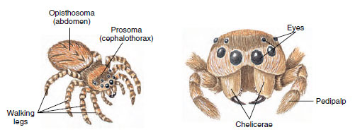
Order Araneae: Spiders
Spiders are a large group of 35,000 species, distributed throughout the world. The spider body is compact: a cephalothorax (prosoma) and abdomen (opisthosoma), both unsegmented and joined by a slender pedicel.
Anterior appendages are a pair of chelicerae (Figure 18-5), which have terminal fangs through which run ducts from poison glands, and a pair of pedipalps having basal parts with which they chew (Figure 18-5). Four pairs of walking legs terminate in claws.
All spiders are predaceous, feeding largely on insects. They effectively dispatch their prey with their fangs and poison. Some spiders chase prey, others ambush them, and many trap them in a net of silk. After a spider seizes prey with its chelicerae and injects venom, it liquefies the tissues with a digestive fluid and sucks the resulting broth into the stomach. Spiders with teeth at the bases of chelicerae crush or chew prey, aiding digestion by enzymes from their mouth.
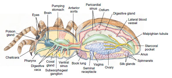
Spiders breathe by means of book lungs or tracheae or both. Book lungs, which are unique to spiders, consist of many parallel air pockets extending into a blood-filled chamber (Figure 18-6). Air enters the chamber by a slit in the body wall. Tracheae make up a system of air tubes that carry air directly to the tissues from openings called spiracles. The tracheae are similar to those in insects but are much less extensive.
Spiders and insects have a unique excretory system of malpighian tubules (Figure 18-6), which work in conjunction with specialized rectal glands. Potassium and other solutes and waste materials are secreted into the tubules, which drain the fluid, or “urine,” into the intestine. Rectal glands reabsorb most potassium and water, leaving behind such wastes as uric acid. By this cycling of water and potassium, species living in dry environments may conserve body fluids, producing a nearly dry mixture of urine and feces. Many spiders also have coxal glands, which are modified nephridia that open at the coxa, or base, of the first and third walking legs.
Spiders usually have eight simple eyes, each with a lens, optic rods, and a retina (Figure 18-6). They are used chiefly for perception of moving objects, but some, such as those of hunting and jumping spiders, may form images. Since a spider’s vision is usually poor, its awareness of its environment depends largely on its hairlike sensory setae. Every seta on its surface, regardless of whether it is actually connected to receptor cells, is useful in communicating some information about the surroundings, air currents, or changing tensions in the spider’s web. By sensing vibrations of its web, a spider can judge the size and activity of its entangled prey or can receive the message tapped out by a prospective mate.
 |
| Figure 18-6 Spider, internal anatomy. |
Web-Spinning Habits: The ability to spin silk is central to a spider’s life, as it is in some other arachnids. Two or three pairs of spinnerets containing hundreds of microscopic tubes run to special abdominal silk glands (Figure 18-6). A scleroprotein secretion emitted as a liquid apparently hardens as a result of being pulled from the spinnerets and forms a silk thread. Spiders’ silk threads are stronger than steel threads of the same diameter and are considered second in strength only to fused quartz fibers. The threads will stretch one-fifth of their length before breaking.
 |
| Figure 18-7 Grasshopper, snared and helpless in the web of a golden garden spider (Argiope aurantia), is wrapped in silk while still alive. If the spider is not hungry, the prize will be saved for a later meal. |
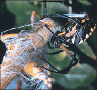
A web used for trapping insects is the use of silk familiar to most people. The kind of net varies among species. Some are simple and consist merely of a few strands of silk radiating out from a spider’s burrow or place of retreat. Others spin beautiful, geometrical orb webs. However, spiders use silk threads for many purposes besides web making. They use them to line their nests; form sperm webs or egg sacs; build draglines; make bridge lines, warning threads, molting threads, attachment discs, or nursery webs; or to wrap their prey securely (Figure 18-7). Not all spiders spin webs for traps. Some, such as wolf spiders, jumping spiders (Figure 18-5), and fisher spiders (Figure 18-8), simply chase and catch their prey.
Reproduction: Before mating, a male spins a small web, deposits a drop of sperm on it, and then picks the sperm up and stores it in special cavities of his pedipalps. When he mates, he inserts his pedipalps into the female genital opening to store the sperm in his mate’s seminal receptacles. A courtship ritual usually precedes mating. A female lays her eggs in a silken net, which she may carry about or attach to a web or plant. A cocoon may contain hundreds of eggs, which hatch in approximately two weeks. Young usually remain in the egg sac for a few weeks and molt once before leaving it. Several molts occur before adulthood.
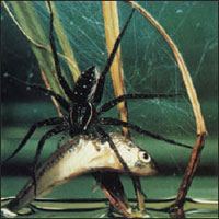 |
| Figure 18-8 Fisher spider, Dolomedes triton, feeds on a minnow. This handsome spider feeds mostly on aquatic and terrestrial insects but occasionally captures small fishes and tadpoles. It pulls its paralyzed victim from the water, pumps in digestive enzymes, then sucks out the predigested contents. |
Are Spiders Really Dangerous? It is truly amazing that such small and helpless creatures as spiders have generated so much unreasoning fear in human minds. Spiders are timid creatures that, rather than being dangerous enemies to humans, are actually allies in the continuing battle with insects. Venom produced to kill prey is usually harmless to humans. Even the most poisonous spiders bite only when threatened or when defending their eggs or young. American tarantulas (Figure 18-9), despite their fearsome size, are not dangerous. They rarely bite, and their bite is not serious.
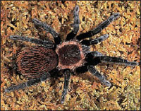 |
| Figure 18-9 A tarantula, Brachypelma vagans. |
There are, however, two genera in the United States that can give severe or even fatal bites: Latrodectus (L. latro, robber, + dektes, biter), and Loxosceles (Gr. loxos, crooked, + skelos, leg). The most important species are Latrodectus mactans, the black widow, and Loxosceles reclusa, the brown recluse. Black widows are moderate to small in size and shiny black, with a bright orange or red “hourglass” on the underside of the abdomen (Figure 18-10A). Their venom is neurotoxic, acting on the nervous system. About four or five of each 1000 bites reported have proved fatal.
Brown recluse spiders are brown and bear a violin-shaped dorsal stripe on their back (Figure 18-10B). Their venom is hemolytic rather than neurotoxic, producing death of tissues and skin surrounding the bite. Their bite can be mild to serious and occasionally fatal.
Some spiders in other parts of the world are dangerous, for example, funnelweb spiders Atrax robustus in Australia. Most dangerous of all are certain ctenid spiders in South America, for example, Phoneutria fera. In contrast to most spiders, these are quite aggressive.
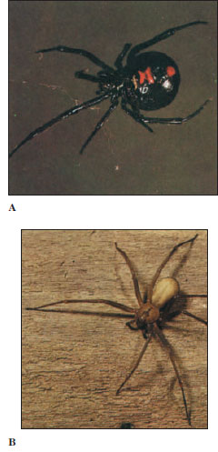 |
| Figure 18-10 A, Black widow spider, Latrodectus mactans, suspended on her web. Note the red “hourglass” on the ventral side of her abdomen. B, Brown recluse spider, Loxosceles reclusa, is a small venomous spider. Note the small violinshaped marking on its cephalothorax. The venom is hemolytic and dangerous. |
Order Scorpionida: Scorpions
Although scorpions are more common in tropical and subtropical regions, some occur in temperate zones. Scorpions are generally secretive, hiding in burrows or under objects by day and feeding at night. They feed largely on insects and spiders, which they seize with their pedipalps and tear up with their chelicerae.
Sand-dwelling scorpions apparently locate prey by sensing surface waves generated by movements of insects on or in the sand. These waves are picked up by compound slit sensilla located on the basitarsal segments of the legs. A scorpion can locate a burrowing cockroach 50 cm away and reach it in three or four quick orientation movements.
Scorpion tagmata are a rather short cephalothorax, which bears the appendages, a pair of large median eyes, and two to five pairs of small lateral eyes; a preabdomen of seven segments; and a long slender post-abdomen, or tail, of five segments, which ends in a stinging apparatus (Figure 18-11A). Their chelicerae are small and three jointed; their pedipalps are large, chelate (pincerlike), and six jointed; and the four pairs of walking legs are eight jointed.
On the ventral side of the abdomen are curious comblike pectines, which are tactile organs used for exploring the ground and for sex recognition. The stinger on the last segment consists of a bulbous base and a curved barb that injects venom. Venom of most species is not harmful to humans but may produce a painful swelling. However, the sting of certain species of Androctonus in Africa and Centruroides (Gr. kenteo, to prick, + oura, tail, + oides, form) in Mexico can be fatal unless antivenin is administered.
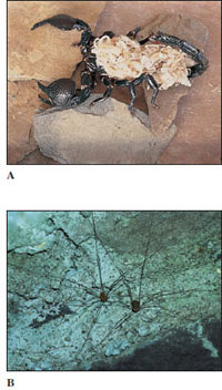
Scorpions perform a complex mating dance, the male holding the female’s chelae and stepping back and forth. He taps her genital area with his forelegs and stings her pedipalp. Finally, he deposits a spermatophore and pulls the female over it until the sperm mass is taken up in the female orifice. Scorpions may be ovoviviparous or truly viviparous; in both cases females brood their young within their reproductive tract. After several months or a year of development, anywhere from 6 to 90 young are produced, depending on the species. The young, only a few millimeters long, crawl onto the mother’s back until after the first molt (Figure 18-11). They mature in about a year.
Order Opiliones: Harvestmen
Harvestmen, often known as “daddy longlegs,” are common in the United States and other parts of the world (Figure 18-11B). These curious creatures are easily distinguished from spiders; their abdomen and cephalothorax are broadly joined, without constriction of a pedicel, and their abdomen shows external segmentation. They have four pairs of usually long, spindly legs, and they can cast off one or more of these without apparent ill effect if they are grasped by a predator (or human hand). The ends of their chelicerae are pincerlike, and they feed much more as scavengers than do spiders.
 |
 |
| Figure 18-11 A, An emperor scorpion (order Scorpionida), Paninus imperator, with young, which stay with the mother until their first molt. B, Harvestmen, Mitopus sp. (order Opiliones). Harvestmen run rapidly on their stiltlike legs. They are especially noticeable during the harvesting season, hence the common name. |
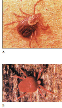
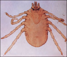
Figure 18-12 A, Wood tick, Dermacentor variabilis (order Acari). Larvae, nymphs, and adults are all parasitic but drop off their hosts to molt to the next stage. B, Red velvet (harvest) mite, Trombidium sp. As with chiggers (Trombicula), only larvae of Trombidium are parasitic. Nymphs and adults are free living and feed on insect eggs and small invertebrates. |
Order Acari: Ticks and Mites
Members of order Acari are without doubt the most medically and economically important group of arachnids. They far exceed the other orders in numbers of individuals and species. Although about 30,000 species have been described, some authorities estimate that from 500,000 to 1 million species exist. Hundreds of individuals of several species of mites may be found in a small portion of leaf mold in forests. They occur throughout the world in both terrestrial and aquatic habitats, even extending into such inhospitable regions as deserts, polar areas, and hot springs. Many acarines are parasitic during one or more stages of their life cycle.
Most mites are 1 mm or less in length. Ticks, which are only one suborder of Acari, range from a few millimeters to occasionally 3 cm. A tick may become enormously distended with blood after feeding on its host.
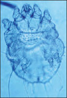
Acarines differ from all other arachnids in having complete fusion of the cephalothorax and abdomen, with no sign of external division or segmentation (Figure 18-12). They carry their mouthparts on a little anterior projection, the capitulum. The capitulum mainly consists of the feeding appendages surrounding the mouth. On each side of their mouth is a chelicera, which functions in piercing, tearing, or gripping food. The form of the chelicerae varies greatly in different families. Lateral to the chelicerae is a pair of segmental pedipalps, which also vary greatly in form and function related to feeding. Ventrally the bases of the pedipalps fuse to form a hypostome, whereas a rostrum, or tectum, extends dorsally over their mouth. Adult mites and ticks usually have four pairs of legs, although there may be only one to three in some specialized forms.
Most acarines transfer sperm directly, but many species use a spermatophore. A larva with six legs hatches from the egg, and one or more eight-legged nymphal stages follow before the adult stage is reached.
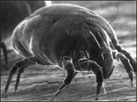 |
| Figure 18-13 Scanning electron micrograph of house dust mite, Dermatophagoides farinae. |
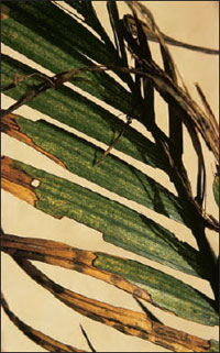 |
| Figure 18-14 Damage to Chamaedorea sp. palm caused by mites of the family Tetranychidae (order Acari). Over 130 species of this family occur in North America, and some are serious agricultural pests. Mites pierce plant cells and suck out contents, giving leaves the mottled appearance shown here. |
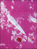
Many species of mites are entirely free living. Dermatophagoides farinae (Gr. dermatos, skin, + phago, to eat, + eidos, likeness of form) (Figure 18-13) and related species are denizens of house dust all over the world, sometimes causing allergies and dermatoses. There are some marine mites, but most aquatic species are found in fresh water. They have long, hairlike setae on their legs for swimming, and their larvae may be parasitic on aquatic invertebrates. Such abundant organisms must be important ecologically, but many acarines have more direct effects on our food supply and health. Spider mites (family Tetranychidae) are serious agricultural pests on fruit trees, cotton, clover, and many other plants. They suck out the contents of plant cells, causing a mottled appearance to the leaves (Figure 18-14), and construct a protective web from silk glands opening near the base of the chelicerae. Larvae of the genus Trombicula are called chiggers or redbugs. They feed on the dermal tissues of terrestrial vertebrates, including humans, and may cause an irritating dermatitis; some species of chiggers transmit a disease called Asiatic scrub typhus. Hair follicle mites, Demodex (Figure 18-15), are apparently nonpathogenic in humans; they infect most of us although we are unaware of them. Other species of Demodex and other genera of mites cause mange in domestic animals. Human itch mites, Sarcoptes scabiei (Figure 18-16), cause intense itching as they burrow beneath the skin.
In addition to disease conditions that they themselves cause, ticks are among the world’s premier disease vectors, ranking second only to mosquitos. They surpass other arthropods in carrying a great variety of infectious agents including apicomplexans, rickettsial, viral, bacterial, and fungal organisms. Species of Ixodes carry the most common arthropod-borne infection in the United States, Lyme disease (see note). Species of Dermacentor (Figure 18-12A) and other ticks transmit Rocky Mountain spotted fever, a poorly named disease because most cases occur in the eastern United States. Dermacentor also transmits tularemia and agents of several other diseases. Texas cattle fever, also called redwater fever, is caused by a protozoan parasite transmitted by cattle ticks Boophilus annulatus (Figure 18-17). Many more examples could be cited.
 |
 |
 |
| Figure 18-15 Demodex folliculorum, human follicle mite. |
Figure 18-16 Sarcoptes scabiei, itch mite. |
Figure 18-17 Boophilus annulatus, a tick that carries Texas cattle fever. |
Support our developers


