The Crocodilia
Crocodiles, the highest living Reptilia, are Lacertilian in form, with long tails and four well-developed limbs, the anterior pair being the shorter, and possessing five complete digits, while the hind-feet are four-toed. With a single exception, the living species have nails on the three preaxial (radial and tibial) digits, so that two digits are without nails on the fore-foot, and one on the hind-foot. The feet are webbed, but the degree to which the web is developed varies greatly. The nostrils are situated at the end of the long snout, and can be closed. The tympanic membranes are exposed, but a cutaneous valve, or earlid, lies above each, and can be shut down over it. All are partially aquatic in habit, and some (the Gavials) are completely so. None of the existing genera are marine, though many ancient Crocodilia inhabited the sea.The dermal armor is composed of scutes covered by epidermic scales of corresponding form. When the armor is complete-as in Caiman and Jacare alone among existing Crocodilia, in Teleosaurus and Stagonolepis among extinct forms-it consists of transverse rows of quadrate bony plates, disposed so as to form a distinct dorsal and ventral shield, separated by soft integument, in the trunk, but united into continuous rings on the tail. The scutes of the same row are united suturally; those of each row overlap their successors, which present smooth facets to receive their under-surfaces. In existing Crocodilia, in the extinct Crocodilus Hastingsiae, and in Stagonolepis, each ventral scute consists of two pieces, a small anterior and a large posterior, united by a suture, The scutes always exhibit a pitted sculpture, and those of the dorsal region are ridged longitudinally, while the ventral scales are always flat. More or fewer dorsal scutes exist in all crocodiles, and those upon the neck sometimes form distinct "nuchal" and "cervical" groups, distinct from the dorsal shield. The dorsal scutes do not always overlap, and the ventral scutes are absent, or incompletely ossified, in most existing Crocodilia.
In these reptiles the vertebral column is always thoroughly ossified, and marked out into distinct cervical, dorsal, lumbar, sacral, and caudal regions. The number of the presacral vertebrae is twenty-four; that of the sacral, two, in all the recent forms, and probably in the extinct genera also. The number of the caudal vertebra varies, but is not less than thirty-five. The number of the cervical, dorsal, and lumbar vertebrae varies; but there are usually nine of the first, eleven or twelve of the second, and four, or three, of the third description.
In existing Crocodilia all the vertebra, except the atlas and axis, the two sacrals, and the first caudal, are procoelous. The majority of the pre-cretaceous Crocodilia have the corresponding vertebrae amphicoelous, the concavities of the centra being very shallow. One genus, Streptospondylus, which is perhaps Crocodilian, has the anterior vertebrae opisthocoelous. It is characteristic of the Crocodilia, that the centra of the vertebrae are united by fibro-cartilages, and that the neurocentral sutures persist for a long time, or throughout life.
The atlas is composed of four pieces, an upper median piece-which is sometimes divided into two, and is developed in membrane apart from the rest-being added to the three pieces found in Lacertilia and Chelonia. A large odontoid bone is closely united to, but not anchylosed with, the anterior flat face of the second vertebra. A pair of elongated, single headed ribs are attached to the inferior piece of the atlas, and another similar pair to the os odontoidum and to the second vertebra, by distinct capitular and tubercular processes. The other cervical vertebrae all possess ribs with distinct and long capitula and tubercula-the latter attached above the neurocentral suture to the neural arch, the former to the centrum below the neurocentral suture. The body of each cervical rib, after the second, and as far as the seventh or eighth, is short, and prolonged in front of, as well as behind, the junction of the capitulum with the tuberculum; and the several ribs lie nearly parallel with the vertebral column, and overlap one another. The ribs of the eighth and ninth cervical vertebrae are longer, and take on more the character of the dorsal ribs, the ninth having a terminal cartilage.
The points to which the capitula and tubercula of the ribs are attached are raised into tubercles; and, by degrees, these become elongated into distinct capitular and tubercular processes, between which, in the third to the ninth vertebrae, the neurocentral suture passes. But in the tenth and in the eleventh vertebrae, the capitular process, which lies nearer the neurocentral suture in the posterior than in the anterior cervical vertebrae, rises upon the body of the vertebra to the level of the neurocentral suture, by which it is traversed, and the tubercular process becomes longer than it. (See Fig. 5, p. 19.) The terminal cartilage is united with the sternum by a sternal rib, which may become more or less completely converted into a cartilage-bone, and is articulated with the vertebral rib.
In the twelfth vertebra a sudden change in the character of the transverse processes takes place. There is no longer a capitular, distinct from a tubercular, process, but one long "transverse process" takes the place of both. A sort of step in the base of this process bears the capitulum of the rib, and answers to the capitular process of the cervical vertebra, while the outer end of the process articulates with the tuberculum of the rib, and represents the tubercular process. The neurocentral suture, in this and the succeeding dorsal vertebrae, lies below the root of the transverse process, which, therefore, is wholly a product of the neural arch. Neither the capitular processes, nor that part of the dorsal transverse process which represents them, have distinct centres of ossification. (Thus, if it be a part of the definition of a "parapophysis," that it is antogenous, there are no parapophyses in the vertebrae of the Crocodilia; and if it be part of the definitation of a "parapophysis" that its arises from the centrum, the dorsal vertebrae of the Crocodilia have no parapophyses.)
In the succeeding dorsal vertebrae the "step" of the transverse process gradually moves outward, until at length it becomes confounded with the tubercular facet, and a corresponding change takes place in the proximal ends of the ribs, in the hindermost of which the distinction between capitulum and tuberculum is lost.
The lumbar vertebrae have long transverse processes which arise from the neural arches, i. e., above the neurocentral suture.
The centra of the two sacral vertebrae have their applied and firmly-united faces flat, their free faces concave; consequently, the first has the anterior face concave and the posterior flat, while the second has the anterior surface flat and the posterior concave. Each sacral vertebra has a strong rib expanded at its distal end; and wedged in at its proximal end, between rough sutural surfaces furnished by the neural arch above and the centrum below.
The first caudal vertebra is biconvex, but all the others are procoelous; those of the anterior moiety of the tail have long ribs fixed in between the neural arches and centra, as in the sacrum, and becoming anchylosed in that position. Chevronbones are attached to the posterior edges of the centra of the vertebrae, except that of the first, and those of the posterior part of the tail.
From seven to nine of the anterior dorsal ribs are united with the sternum by sternal ribs, the form of which varies a good deal in different Crocodilia, being sometimes narrow, sometimes broad and flattened. An elongated plate of cartilage, which may be partially converted into cartilage-bone, is attached to the hinder margin of several of the most anterior ribs, above the junction between the ossified and the cartilaginous part of the vertebral rib. (Fig. 5, P.u.) These are the so-called "uncinate processes," which also exist in Hatteria, and reappear in Birds.
The sternum consists of a rhomboidal plate of cartilagebone, with the posterolateral edges of which two pairs of sternal ribs articulate. The posterior angle of the plate is continued into a median prolongation, which, at length, divides into two curved divergent cornua. From five to seven pairs of sternal ribs are united with the prolongation and its cornua. A long and slender interclavicle lies in a groove of the middle of the ventral face of the rhomboidal part of the sternum.
In the ventral wall of the abdomen, superficial to the recti muscles, lie seven transverse series of membrane-bones, which are termed "abdominal ribs;" though it must be recollected that they are quite distinct from true ribs, and rather correspond with the dermal ossicles of the Labyrinthodonta. Each series is composed of four elongated and more or less curved ossicles, pointed at each end, and so disposed that inner ends of the inner pair meet at an angle, open backward in the middle line, while their outer ends overlap the inner ends of the outer pair. The most posterior of these ossicles are stronger than the others, and are closely connected with the pubic cartilages.
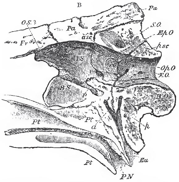 |
| Fig. 77. - Longitudinal and vertical section of the hinder part of the skull of a Crocodile; Eu, Eustachian tube: P N, posterior nares; P, pituitary fossa. |
1. There is an interorbital septum, and the presphenoidal and orbitosphenoidal regions remain cartilaginous, or very incompletely ossified.
2. All the bones of the skull (except the mandible, stapes, and hyoid) are firmly united by sutures, which-persist throughout life.
3. There are large parotic processes. Both the upper and the lower temporal arcades are completely ossified, and formed by post-frontal, squamosal, jugal, and quadrato-jugal bones; supra-temporal, lateral-temporal, and post-temporal fossae are formed, as in the Lacertilia, though their relative sizes are very diiferent.
4. The maxillary and the palatine bones develop palatine plates, which unite suturally in the middle line, and separate the nasal passages from the cavity of the mouth, as in Mammalia; and in all existing Crocodiles, but not in Teleosaurus or Belodon, the pterygoids are also modified in the same way (as in Mymercophaga among Mammals), so that the posterior nares are situated very far back beneath the base of the skull.
5. In consequence of the development of these palatine plates of the maxillary and palatine bones, the two vomers are, in most Crocodiles, invisible upon the under-surface of the bony roof of the mouth.
6. There are larger alisphenoids, but the orbitosphenoids are absent or radimentary.
7. There is no parietal foramen.
8. The quadrate bone is very large, and fixed immovably to the walls of the skull, as in the Chelonia; and, as in the latter, the pterygoid bone is firmly connected with the base of the skull, and united only with the upper and inner surface of the quadrate bone.
9. The pterygoid sends down a large free process, against the broad outer edge of which the inner surface of the mandible plays.
10. The tympanic cavity is completely bounded by bone. The prootic and opisthotic (which is united with the ex-occipital) form its inner walls, the quadrate its outer wall, the squamosal and post-frontal its roof, and the quadrate, the basi-occipital, and basisphenoid its floor. The two tympana are placed in communication with the cavity of the mouth by three canals-one large, opening in the middle line; and two smaller ones at the sides, on the base of the skull, behind the posterior nares. The large canal passes up between the basisphenoid and basi-occipital, and divides between those bones into a right and left lateral canal. Each lateral canal subdivides into an anterior branch, which traverses the basisphenoid, and a posterior, which passes up in the basi-occipital. The posterior branch receives the narrow lateral canal of its side (which runs vertically up to it), and then opens into the posterior part of the floor of the tympanum. The anterior branch opens into its anterior wall.
The tympanic cavities of embryonic Crocodiles communicate with the mouth by wide and simple apertures, and the complicated arrangement of canals just described results from the great downward development of the basisphenoid and basi-occipital, and their encroachment upon these apertures on the inner side, while the quadrate bone narrows them on the outer.
In adult Crocodilia, air-passages extend from each tympanum to that of the opposite side, through the bones which form the roof of the posterior region of the skull. On the other hand, they excavate the quadrate bone, whence the air passes through a membranous tube into the hollow articular piece of the mandible. The hyoidean apparatus is greatly simplified, consisting only of a broad plate of cartilage, which may become partially ossified, and two ossified cornua which are not directly connected a with the skull. A minute styliform cartilage, which lies in close proximity with the portio dura, on the upper part of the posterior face of the quadrate bone, represents the stylohyal, or proximal end of the hyoidean arch.
The pectoral arch has no clavicle, and the coracoid has no distinct epicoraooidal element, nor any fontanelle. The carpus consists proximally of two elongated and somewhat hour-glassshaped bones, articulated respectively with the radius and the ulna. The radial is the larger, and is partially articulated with the ulna. Behind these, and directed transversely, lies another curved ossification, the upper concave face of which articulates with the ulna. It is united with the latter bone on the one hand, and with the fifth metacarpal, on the other, by strong ligaments, and represents a pisiform bone. Distally, there lies on the ulnar side the so-called lenticular bone, an oval ossicle interposed between the ulnar proximal carpal and the second, third, fourth, and fifth metacarpals, the last three of which it supports altogether. On the radial side, a disk of cartilage, which never becomes completely ossified, is connected by ligament with the lenticulare, and is interposed between the radial proximal bone and the head of the metacarpal of the pollex. From the ulnar side of the head of this bone a cartilaginous ligamentous band proceeds, over the head of the second metacarpal, to the radial side of the lenticulare.
The three radial digits are much stronger than the two ulnar, and the numbers of the phalanges are 2, 3, 4, 4, 3, counting from the radial to the ulnar side.
The pelvis (Fig. 78, C) possesses large ilia, which are firmly united with the expanded ends of the strong ribs of the sacrum. The ischium unites with its fellow in a median ventral symphysis, and, with the ilium, forms almost the whole of the acetabulum.
The pubes take hardly any share in the formation of the latter cavity in the adult. Their axes are directed forward and inward, and they coalesce in the middle line; but as the inner, or median, moiety of each pubis remains cartilaginous, or imperfectly ossified, the bones, in imperfectly prepared skeletons, appear as if they formed no symphysis.
The tarsus presents, proximally, an astragalo-navicular bone and a calcaneum, which are less closely united than in the Lizards. The latter bone has a large calcaneal process on Its posterior face, the Crocodile being the only Sauropsid vertebrate in which such a process is developed (Fig. 78, C. Ca.).
Two rounded distal tarsal bones, of which the fibular is much the larger, lie between the calcaneum and the third, fourth, and rudimentary fifth, metatarsals. A thin plate of cartilage is interposed between the distal end of the astragalonavicular and the second metatarsal, and unites with the head of the first metatarsal.
As in the manus, the three, pre-axial, clawed, digits are stronger than the others. The fifth is represented only by an imperfect metatarsal. The numbers of the phalanges are 2, 3, 4, 4, counting from the tibial to the fibular side.
In the Crocodilia the teeth are confined to the premaxillae, maxillae, and dentary part of the mandible. They are simple in structure, have large pulp-cavities, are lodged in distinct alvcoli, and are replaced by others developed upon their inner sides. The development of the new tooth causes absorption of the inner wall of the base of the old one, and the replacing tooth thus comes to lie within the pulp-cavity of its predecessor. The teeth vary much in shape, having either long, curved, and acute, or short and obtuse, or almost globular and straight, crowns. Very often they possess sharp anterior and posterior edges, which may be finely serrated.
The Crocodilia are to be found in the rivers of all continents and the larger islands in the hotter parts of the world. None of the existing species are truly marine, though many of the extinct species were. They are first known to occur in strata of Triassic age, and abound, under forms which differ but little from some of those which now exist, in the Mesozoic and Cainozoic formations.
They may be divided into the following groups:
- With procoelous presacral vertebrae, and posterior nares bounded below by the pterygoids. (All existing Crocodilia, and the fossil forms of cretaceous and later formations, are included in this division.)
- The nasals enter into the formation of the nasal aperture.
- The head short and broad. The teeth very unequal; the first and fourth of the mandibles biting into pits of the upper jaw. The premaxillo-maxillary suture straight or convex forward. The mandibular symphysis not extending beyond the fifth tooth, and the splenial element not entering into it. The cervical scutes distinct from the tergal. 1. Alligaloridae.
- The head longer. The teeth unequal. The first mandibular
tooth biting into a fossa; the fourth, into a groove, at the
side of the upper jaw. The premaxillo-maxillary suture
straight or convex backward. The mandibular symphysis
not extending beyond the eighth tooth, and not involving
the splenial elements. The cervical scutes sometimes distinct
from the tergal, sometimes united with them.
2. Crocodilidae.
Crocodilus. Mecistops.
- The nasals are excluded from the external nasal aperture. The head very long; the teeth subequal. Both the first and the fourth mandibular teeth bite into grooves in the margin of the upper jaw. The premaxillo-maxillary suture acutely angulated backward. The mandibular symphysis extends to at least the fourteenth tooth, and the splenials enter into it. The cervical and tergal seutes form a continuous series. 3. Guvialidae.
- With the presacral vertebrae amphieoelons (the anterior vertebrae sometimes opisthocoelous (?) ); and the posterior nares bounded by the palatines, the pterygoids not being united below. (All these Crocodiles are extinct and pre-cretaceous.)
Alligator. Caiman, Jacare.
Rhynchosuchus. Gavialis.
- With the external nares terminal.
4. Telesauridae.
Telesaurus. Goniopholis.
Streptospondylus. Stagonolepis. Galesaurus (?). - With the external nares on the upper part of the base of the snout
near the orbits.
5. Belodontidae.
Belodon.
There is a large number of extinct Reptilia which resemble the Crocodilia in the characters of their pre-sacral vertebrae, but differ from them, and resemble Lacertilia Chelonia, or Birds, in other respects.
These are the Dicynodontia, the Ornithoscelida, and the Pterosauria.
Support our developers


