Protozoa and Animal Parasites
Medical parasitology is concerned with the study and identification of the pathogenic protozoa and helminths (worms) that cause the parasitic diseases of humans and animals.Protozoa
Protozoa are the largest of the unicellular true microorganisms. They are classified in the Kingdom Protista although their name implies that they were the forerunners of the animal kingdom (proto = first; zoa = animal).
The basic structures of all protozoa include a nucleus well defined by a nuclear membrane, lying within cytoplasm that is enclosed by a thin outer cell membrane. Other specialized structures, such as cilia or flagella (see colorplate 49) for locomotion or a gullet for food intake, vary with different types of protozoa. Six major groups of protozoa are distinguished on the basis of their locomotory structures or their reproductive mechanisms (see fig. 32.1).
Amebae: Simple ameboid forms. Move by bulging and retracting their cytoplasm in any direction. Major pathogen is Entamoeba histolytica(see colorplate 50).
Ciliates: Move by rapid beating of cilia (fine hairs) that cover the cell membrane. Balantidium coli is a protozoan ciliate that may cause human disease.
Flagellates: Possess one or more flagella that give them a lashing motility. Giardia lamblia (see colorplate 51), Trichomonas vaginalis (see colorplate 49), and the trypanosomes are the major pathogens in this group.
Apicomplexa: No special structures for locomotion (some immature forms have ameboid motility). Reproductive cycle includes both immature and mature forms (later called sporozoites). Toxoplasma gondii and Plasmodium species (see colorplate 52), which are the malarial parasites, are the representative pathogens in this group.
Coccidia: Represent a subphylum of the Apicomplexa. Coccidia have a complex life cycle in which all stages of parasite development are intracellular. Major genera include Cryptosporidium (see colorplate 53), Cyclospora, and Isospora.
Microspora: Includes a large group of obligate, intracellular protozoa that produce spores. These protozoa are classified in more than 100 genera and 1,200 species, collectively called microsporidia. Major genera causing human disease are Enterocytozoon, Encephalitozoon, Nosema, and Pleistophora.
Diagramatic examples of the amebae, ciliates, flagellates, and the Apicomplexa are shown in figure 32.1.
As indicated, species from each of these protozoan groups are associated with human diseases. Some of them are carried into the body through the gastrointestinal tract (in contaminated food or water or by direct fecal contamination of objects placed in the mouth), localize there, and produce diarrhea or dysentery. Others are carried by arthropods, which inject them into the body when they bite. This group of protozoa then infects the blood and other deep tissues. The pathogenic protozoa are summarized in table 32.1.
It should also be noted that some of the intestinal protozoa may live normally in the bowel without causing damage under ordinary circumstances. Some flagellated protozoa frequently are found on the superficial urogenital membranes and sometimes are troublesome when they multiply extensively and irritate local tissues.
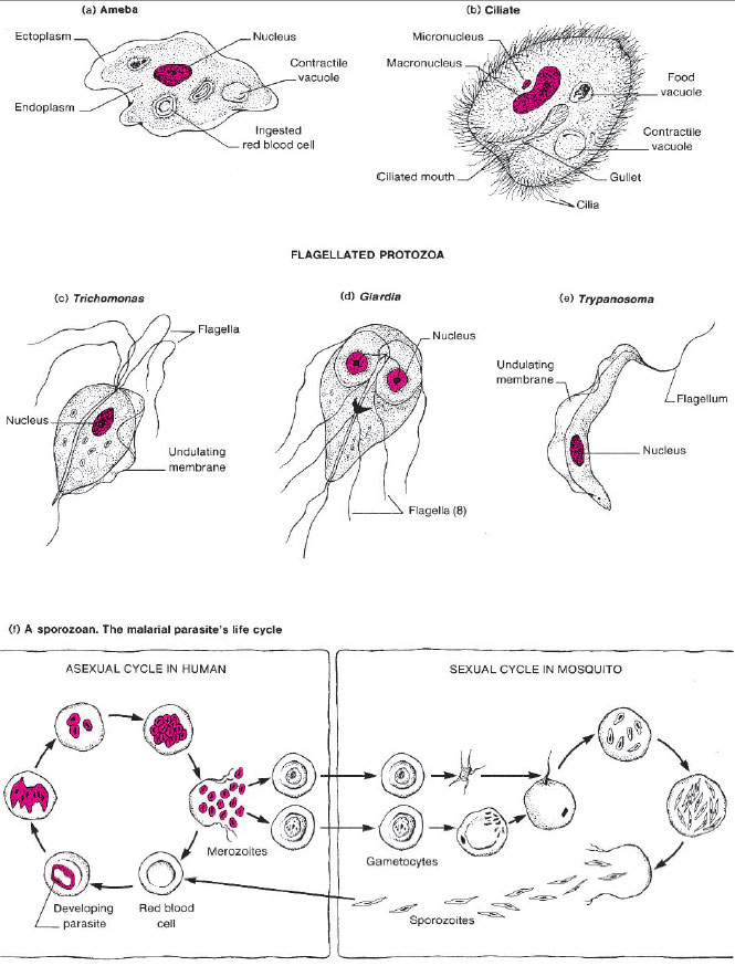 |
| Figure 32.1 Diagrams of four types of protozoa. (a) An active ameba. (b) A ciliated protozoan (Balantidium coli), (c), (d), and (e) Three types of flagellated protozoa. (f) Developmental stages of the malarial parasite, a sporozoan (Plasmodium species). |
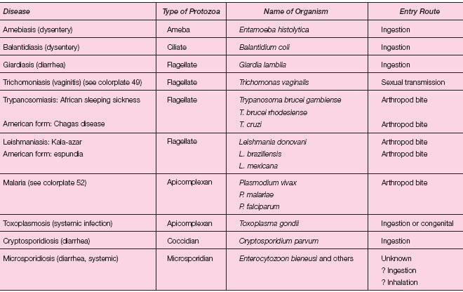 |
| Table 32.1 Pathogenic Protozoa |
*(see colorplate 52)
Other amebae live freely in the environment, in soil and water. Under special circumstances, some of these organisms can infect humans. Members of the genus Naegleria inhabit freshwater ponds, lakes, and quarries. When people dive or swim in water containing the amebae, the organisms can be forced up with water through the thin nasal passages, directly into the central nervous system to cause an almost universally fatal meningoencephalitis (affects both meninges and brain). Acanthamoeba species (see fig. 32.2) are associated with corneal infections in persons whose contact lenses or contact lens care solutions become contaminated by the amebae. To avoid infection these lenses and care solutions must be kept meticulously clean. Corneal transplant is usually required for patients with Acanthamoeba eye infection.
Parasitic Helminths
Helminths, or worms, are soft-bodied invertebrate animals. Their adult forms range in size from a few millimeters to a meter or more in length, but their immature stages (eggs, or ova, and larvae) are of microscopic dimensions. Relatively few species of helminths are parasitic for humans, but these few are widely distributed. It has been estimated that 30% of the earth’s human inhabitants harbor some species of parasitic worm.
There are two major groups of helminths: the roundworms, or nematodes, and the flatworms, or platyhelminths. The latter are again subdivided into two groups: the tapeworms (cestodes) and flukes (trematodes). A summary of the major characteristics of these groups is given here.
Roundworms (Nematodes): Roundworms are cylindrical worms with bilateral symmetry. Most species have two sexes, the female being a copious egg producer. These ova hatch into larval forms that go through several stages and finally develop into adults. In some instances, the eggs of these worms are infective for humans when swallowed. In the intestinal tract they develop into adults and produce local symptoms of disease. In other cases, the larval form, which develops in soil, is infective when it penetrates the skin and is carried through the body, finding its way finally into the intestinal tract where the adults develop. In the case of Trichinella (the agent of trichinosis), the larvae are ingested in infected meat, but penetrate beyond the bowel and become encysted in muscle tissue. One group of roundworms, the filaria, are carried by arthropods and enter the body by way of an insect bite. (See table 32.2.)
Flatworms (Platyhelminths): Flatworms are flattened worms that also show bilateral symmetry. Some are long and segmented (tapeworms); others are short and nonsegmented. Most are hermaphroditic.
Tapeworms (Cestodes): Tapeworms are long, ribbonlike flatworms composed of individual segments (proglottids), each of which contains both male and female sex organs. The tiny head, or scolex, may be equipped with hooklets and suckers for attachment to the intestinal
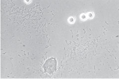 |
| Figure 32.2 Acanthamoeba trophozoite (bottom center) and cysts (refractile objects at top right) isolated from the contact lens of a patient who required a corneal transplant because of the infection. The tiny objects throughout the background are cells of Escherichia coli on which the amebae feed when grown in culture. |
wall. The whole length of the tapeworm, the strobila, may have only three or four proglottids or several hundred. Eggs are produced in the proglottids (which are then said to be gravid) and are extruded into the bowel lumen. Often the gravid proglottids break away intact and are passed in the feces. All tapeworm infections are acquired through ingestion of an infective immature form, in most cases larvae encysted in animal meat or fish (e.g. Diphyllobothrium latum, see colorplate 54). Usually development into adult forms occurs in the intestinal tract, and the tapeworm remains localized there. In one type of tapeworm infection, echinococcosis, the eggs are ingested, penetrate out of the bowel, and develop into larval forms in the deep tissues (see colorplate 55).
Flukes (Trematodes): Some flukes are short, ovoid or leaf-shaped, and hermaphroditic; others are elongate, thin, and bisexual. The flukes are not segmented. They are usually grouped according to the site of the body where the adult lives and produces its eggs, that is, blood, intestinal, liver, and lung flukes. Some of these infections are acquired through the ingestion of larval forms encysted in plant, fish, or animal tissues. In others, a larval form (swimming freely in contaminated water) penetrates the skin and makes its way into deep tissues.
Table 32.2 summarizes the important helminths that cause disease in humans.
Laboratory Diagnosis
Almost all parasitic diseases, whether intestinal or extraintestinal, are diagnosed by finding the organism in appropriate clinical specimens, usually by microscopic examination. Intestinal infections are generally limited to the bowel, and therefore, fecal material is the specimen of choice. In extraintestinal infections, the diagnostic stage of the parasite may be found in blood, tissue, or exudates, so that these specimen types must be examined. With rare exceptions, such as extraintestinal amebiasis and toxoplasmosis, routine serological tests have no application in the diagnosis of parasitic diseases.
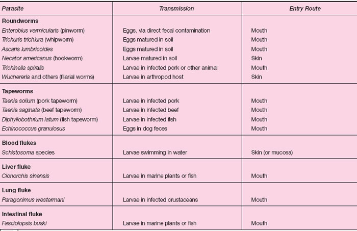 |
| Table 32.2 Important Helminths of Humans |
Intestinal Parasitic Infections
Protozoa or helminths may cause intestinal parasite infections. The laboratory diagnosis of these diseases depends almost exclusively on finding the diagnostic stage(s) in fecal material. If stool samples cannot be examined immediately after passage, a portion of the stool must be placed in a stool collection kit with a special preservative to maintain the structural integrity and morphology of the diagnostic cysts, eggs, or larvae. There is no one perfect stool preservative and the choice usually depends on the laboratory that performs the analysis.
Once a stool is received by the laboratory, the ova and parasite (O&P) examination may consist of any combination or all three of the following techniques: direct wet mount, concentration, and permanent stained smear. Each technique is designed for a particular purpose. Traditionally, the direct examination is used to detect protozoan motility. Since most laboratories use a stool preservative that kills protozoa, direct wet-mount examinations for this purpose are not routinely performed. Instead, the direct wet-mount exam may be used to screen for cysts and eggs that may be present in large numbers in the fecal sample.
Fecal concentration procedures allow for the detection of small numbers of organisms that may be missed when only a direct smear is examined. There are two types of concentration procedures: sedimentation and flotation. Both are designed to separate protozoan cysts and oocysts, microsporidian spores, and helminth eggs and larvae from fecal debris by centrifugation (sedimentation) or differences in specific gravity (flotation).
Stained smears can also be prepared from fecal samples to allow for the improved detection and identification of intestinal protozoa. These slides serve as a permanent record of the organism identified and may be used for teaching purposes as well. Three stains commonly used for the detection of intestinal parasites are the trichrome, iron-hematoxylin, and modified acid-fast stains.
Intestinal Protozoa: The protozoa that parasitize the human intestinal and urogenital systems belong to five major groups: amebae, flagellates, ciliates, coccidia, and microsporidia. With the exception of the flagellate Trichomonas vaginalis (an important cause of vaginitis, see colorplate 49) and microsporidia of the genera Pleistophora, Nosema, and Encephalitozoon, all of these organisms live in and may cause disease of the intestinal tract.
Intestinal helminths: Intestinal helminths are usually diagnosed by the microscopic detection of their eggs or larvae in feces. Characteristics used in identification include size, shape, thickness of shell, special structures of the shell (mammillated covering, operculum, spine, knob) and the developmental stage of egg contents (undeveloped, developing, embryonated). Figure 32.3 shows the relative sizes and comparative morphologies of representative helminth eggs.
Extraintestinal Parasitic Infections
Blood and Tissue Protozoa: Among the protozoa that parasitize human blood and tissue, malaria is detected most frequently in the United States. The laboratory diagnosis of malaria is made by examining blood smears collected from the patient. Blood smears are stained with Giemsa or Wright stain, the common stains also used to examine blood films for hematological parameters. These stains help distinguish the various diagnostic stages and allow for the identification of Plasmodium species (see colorplate 52). Of the four human malarial parasites, Plasmodium vivax and Plasmodium falciparum account for more than 95% of infections, with P. vivax responsible for about 80% of these. Identification of malarial parasites to the species level is important for establishing the prognosis of the disease and predicting the likelihood of drug resistance. Many strains of P. falciparum are now resistant to chloroquine, the drug of choice for treatment. Other more exotic and far less common blood and tissue protozoan diseases seen in the United States are leishmaniasis and trypanosomiasis. These infections, as well as malaria, are almost universally imported into the United States by persons arriving from countries where the parasitic agents are endemic.
Toxoplasma gondii is a tissue protozoan that is an established cause of congenital disease. More recently, toxoplasmosis has been recognized as a cause of central nervous system disease in HIV-infected patients. The diagnosis of toxoplasmosis often depends on the detection or recovery of the organism from tissue biopsy material, CSF specimens, or buffy coat of blood (the white blood cell layer that forms between the erythrocytes and plasma when anticoagulated blood is lightly centrifuged). In general, however, such specimens do not reveal the parasites, even in the presence of active disease. Therefore, serological tests are recommended in all suspected cases of toxoplasmosis.
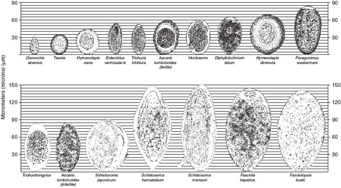 |
| Figure 32.3 Relative sizes and comparative morphologies of representative helminth eggs. |
Tissue Helminths: A large number of helminthic parasites, including nematodes, flukes, and tapeworms, live in human tissues as adults or larvae. Diagnosis of infections caused by them often depends on the identification of the parasite’s reproductive products discharged in blood, feces, or other body fluids or, in the case of larval parasites, on the recovery from or detection of the parasite itself in tissue.
Some of the more common tissue helminths are listed here for your review. The nematodes include the filarial worms Wuchereria bancrofti, Brugia malayi, Onchocerca volvulus, and Loa loa. Strongyloides species (a cause of cutaneous larva migrans), Toxocara canis (the cause of visceral larva migrans), and Trichinella spiralis (the cause of trichinosis) are nematodes that cause disease in the United States. The trematodes include the liver flukes (Fasciola hepatica, Clonorchis sinensis, and Opisthorchis viverrini), lung flukes (Paragonimus westermani), and the blood flukes (Schistosoma species). Finally, are the cestodes or tapeworms, some of the more common of which include Taenia solium (the cause of cysticercosis), Diphyllobothrium latum (the fish tapeworm, see colorplate 54), and Echinococcus granulosus and Echinococcus multilocularis (the causes of hydatid cyst disease, see colorplate 55).
Except where noted, people with tissue helminth diseases become infected outside of the United States. Because of the current ease and frequency of global travel, however, microbiologists throughout the world must become familiar with the laboratory diagnosis of these infections.
Prepared slides and demonstration material will be studied in this exercise.
| Purpose | To study the microscopic morphology of some protozoa and parasitic helminths, and to learn how parasitic diseases are diagnosed |
| Materials | Prepared slides of protozoa Prepared slides of helminth adults, eggs, larvae Projection slides if available |
Procedures
- Examine the prepared slides, audiovisual or reading material, and make drawings of different forms of protozoa and helminths.
- Review demonstration material and assigned reading on the transmission and localization of parasites and complete the table provided under Questions.
Results
Draw each type of organism listed:
- An ameba
- A ciliated protozoan
- A flagellated protozoan
- A protozoan found in blood
- An adult roundworm
- An adult tapeworm
Support our developers


