Frustule
This structure is present only in the Bacillariophyceae (Heterokontophyta). The frustule is an ornate cell membrane made of amorphous hydrated silica, which displays intricate patterns and designs
unique to each species. This silicified envelope consists of two overlapping valves, an epitheca
and a slightly smaller hypotheca. Each theca comprises a highly patterned valve and one or
more girdle bands (cingula) that extend around the circumference of the cell, forming the region
of theca overlay. Extracellular organic coats envelop the plasma membrane under the siliceous frustule. They exist in the form of both thick mucilaginous capsules and thin tightly bound organic
sheaths. The formation of the frustule has place in the silica deposition vesicles, derived from the
Golgi apparatus, wherein the silica is deposited. The vesicles eventually secrete their finished
product onto the cell surface in a precise position.
Diatoms can be divided artificially in centric and pennate because of the symmetry of their frustule. In centric diatoms, the symmetry is radial, that is, the structure of the valve is arranged in reference to a central point (Figure 2.10). However, within the centric series, there are also oval,
triradiate, quadrate, and pentagonal variation of this symmetry, with a valve arranged in reference
to two, three, or more points. Pennate diatoms are bilaterally symmetrical about two axes, apical
and trans-apical, or only in one axis, (Figure 2.11); some genera possess rotational symmetry. Valves of some pennate diatoms are characterized by an elongated fissure, the raphe, which can be placed centrally, or run along one of the edges. At each end of the raphe and at its center there are thickenings called polar and central nodules. Addiction details in the morphology of the frustule are the stria, lines composed of areolae, and pores through the valve that can go straight through the structure, or can be constricted at one side. Striae can be separated by thickened areas called costae. Areolae are passageways for the gases, nutrients exchanges, and mucilage secretion for movement and attachment to substrates or other cells of colony. Other pores, also known as portules, are present on the surface of the valve.
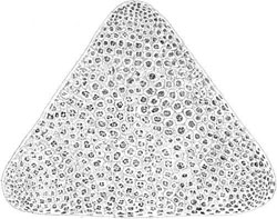
FIGURE 2.10 Triceratium sp., a centric diatom.
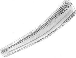
FIGURE 2.11 Rhoicosphenia sp., a pennate diatom.
There are two types of portules: fultoportulae (Figure 2.12) found only in the order Thalassiosirales and rimoportulae (Figure 2.13), which are universal. The structure of the fultoportulae is an external opening on the surface of the valve extended or not into a protruding structure (Figure 2.12). The other end penetrates the silica matrix and is supported with two to five satellite pores. The portules function in the excretion of several materials, among them are b-chitin fibrils. These fibrils are manufactured in the conical invaginations in the matrix, under the portule. This may be the anchoring site for the protoplast. The rimoportula is similar to the fultoportulae, except that it has a simpler inner structure. The rimoportula does not have satellite pores in the inner matrix. However, the rimoportula does have some elaborate outer structures that bend, have slits, or are capped. Sometimes the valve can outgrow beyond its margin in structures called setae that help link adjacent cells into linear colonies as in Chaetoceros spp., or possess protuberances as in Biddulphia spp. that allow the cells to gather in zig-zag chains (Figure 2.14). In other genera such as Skeletonema the valve presents a marginal ridge along its periphery consisting of long, straight spines, which make contact between adjacent cells, and unite them into filaments. Some genera also possess a labiate process, a tube through the valve with internally thickened sides that may be flat or elevated.
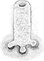
FIGURE 2.12 Fultoportula of Thalassiosira sp.
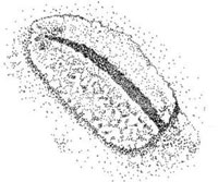
FIGURE 2.13 Rimoportula of Stephanodiscus sp.
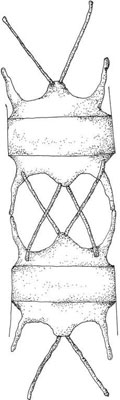
FIGURE 2.14 Cells of Biddulphia sp.
Diatoms are by far the most significant producer of biogenic silica, dominating the marine
silicon cycle. It is estimated that over 30 million km
2 of ocean floor are covered with sedimentary deposits of diatom frustules.


