Microbiology Methods / Basic Techniques of Biotecnologies
Preparing a Wet Mount
| Purpose |
To observe bacteria in a simple wet mount and determine their motility |
| Materials |
24-hour broth culture of Proteus vulgaris mixed with a light suspension of yeast cells
24-hour broth culture of Staphylococcus epidermidis mixed with a light suspension of yeast cells
2 microscope slides
Several cover glasses
Capillary pipettes and pipette bulbs
China-marking pencil or permanent marking pen
Clear nail polish (optional) |
Procedures
-
Using a pipette bulb, aspirate a small amount of the Proteus culture with a capillary pipette and place a small drop on a
clean microscope slide (fig. 3.2, step 1).
-
Carefully place a clean cover glass (see Experiment 3.1, procedure 1) over the drop, trying to avoid bubble formation (fig.
3.2, step 2). The fluid should not leak out from under the edges of the cover glass. If it does, wait until it dries before
sealing.
-
If you examine the slide immediately, you need not seal the coverslip. Otherwise, seal around the edges of the coverslip
with a thin film of clear nail polish (fig. 3.2, step 3). Be certain the nail polish is completely dry before examining the slide
under the microscope.
-
Examine the preparation in the same manner as in Experiment 3.1, following procedures 6 through 10. Instead of
focusing on the edge of the drop, however, you may find it helpful to focus first on the left-hand edge of the coverslip.
-
Make a wet-mount preparation of the Staphylococcus culture, following the same procedures just described.
|
|
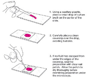 |
| Figure 3.2 Wet-mount preparation. |
|
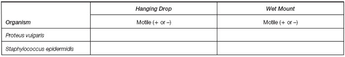
Results
- If you have not performed a hanging drop as in Experiment 3.1, make drawings in the circles on page 28 according to the
directions in the results for that exercise.
- If you have performed Experiment 3.1, complete the chart.
Support our developers

More in this section