Liverworts
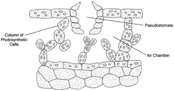 |
| Figure 24-1 Marchantia gametophyte thallus. |
Murchantia is an example of a thallose liverwort. The gametophytet hdus is dichotymously branched and usually approximately two centimeters wide and four to six centimeters long. The sporophytes are nearly microscopically sized. Marchantia is found in wet places, flattened on the soil and anchored by rhizoids extending from its underside. When one examines the thallus with a lens, small openings can be seen. These pores open to chambers immediately below, where columns of cells extend up from the floor of the chamber. These cells have chloroplasts and are active in photosynthesis. Carbon dioxide, which is used in the photosynthetic process, drifts into the chamber through the pore, and oxygen, a product of photosynthesis, drifts outward. The pores may be called pseudostomates, pseudo because they are not considered to be the same as or related to the stomates of more advanced plants.
Three kinds of appendages are found on the underside of the thallus: peg rhizoids, which have internal, peglike structures on the cell wall and function in absorption; smooth rhizoids, which also function in absorption as well as anchor the plant to the soil; and scales, which have no known function but may be protective.
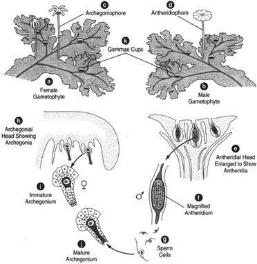 |
| Figure 24-2 Marchantia gametophyte generation. (a) The female gametophyte. (b) The male gametophyte. (c) An archegoniophore at the tip of which are archegonia under the lobes. (d) An antheridiophore bearing antheridia at the tip. (e) An enlarged antheridial head, showing antheridia. (f) A magnified antheridium. (g) A sperm cell. (h) Archegonial head, showing archegonia on the under side of the lobes. (i) An immature archegonium. (j) Amature archegonium. (k) A gemma cup bearing gemmae (agents of a sexual reproduction). |
The sexes are separate. On the female gametophyte, special stalks called archegoniophores rise up from the surface of the thallus. Upon maturity, the archegoniophores have fingerlike lobes at the upper end. Archegonia grow on the undersides of these fingerlike lobes. Each archegonium is a flask-shaped structure bearing an egg cell in its bottom portion, the venter. When the archegonium is quite young, there are cells in the neck portion. These cells must break down and dissolve away as the archegonium matures to allow a passageway for sperm cells.
The male gametophyte also produces stalks, called antheridiophores. Antheridiophores have a different appearance at the upper end than do archegoniophores. The upper end of an antheridiophore is a flat, disc-shaped head on the upper surface of which antheridia are borne. When the antheridia mature, they break open and release large numbers of biflagellated sperm cells. Because the sexes are separate and the gametes are produced at the tops of special stalks, the way whereby sperm reach the egg cells is perplexing. One supposition is that a rain drop striking the tip of an antheridiophore causes the sperm to be catapulted to their destinations. Another is that they are carried by insects. Yet another is that the sperm have great enough endurance to swim down the length of the antheridiophore, across a certain expanse of thallus, up the length of a neighboring archegoniophore, and arrive at the archegonia to fulfill their purpose.
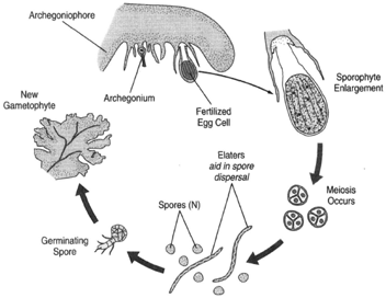 |
| Figure 24-3 Marchantia sporophyte generation. At left, an archegoniophore bearing an archegonium: The egg cell has been fertilized, and the young sporophyte is growing. At upper right, an enlargement of the sporophytes, howing spore mother cells in the capsule. Just below, meiosis occurs. At bottom, a spore germinates to produce a new ametophyte. |
The egg cell is fertilized in the venter of the archegonium, where the sporophyte develops: The sporophyte remains attached to the gametophyte (in a sense, parasitic upon it) and microscopically sized. The sporophyte generation is diploid. It develops a foot, which grows through the base of the archegonium and anchors the sporophyte to the tissue of the gametophyte. A short seta and a capsule in which spores and elaters develop also form. Spore mother cells (diploid cells destined to undergo meiosis and produce haploid spores) are in the capsule. Upon maturity, the capsule splits open, and the spores and elaters are liberated. The elaters are hygroscopic filaments, coiling and uncoiling under the influence of varying conditions of humidity. These movements aiind the dispersal of spores.
Asexual reproduction is achieved either by the thallus fragmenting through decay or an enchanting evolutionary wonder called gemmae. Gemmae are smalcl lusters of cells that are scarcely visible without a lens and rest in a minute basket on the upper surface of the thallus. This little basket is called the gemma cup, and the little, egglike clusters of cells are the gemmae. The gemmae can be dislodged and, falling on favorable soil, grow into new Marchantia plants. The method is entirely vegetative. There is no alternation of generations with gemmae.
Support our developers


