Life Cycle
Now consider flowering plants. The conspicuous generation is the sporophyte. mowers are apart of the sporophyte generation. Flowers are composed of modified leaves arranged in several whorls. The innermost whorl constitutes what is generally known as the female part. This raises an apparent contradiction, however. If the plant is a sporophyte, it seems contradictory to indicate it having female parts. As will be seen, not only is this contradictory, it is also acceptable.The innermost whorl is called the pistil. It possesses the stigma (at its upper end); the long, slender style; and the ovary containing ovules (at the base). The ovules become seeds. Proceeding from the innermost whorl outward, the next whorl of flower parts are the stamens, composed of filaments and anthers. The anthers bear pollen grains. Next come the petals, which compose the corolla. The outermost whorl, the sepals, compose the calyx.
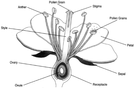 |
| Figure 28-1 A generalized flower. |
Figure 28-2 shows a developing ovule. It is composed of many cells, one of which has a destiny different from that of the others. This cell is the megaspore mother cell, whose destiny is to undergo meiosis to produce a tetrad of haploid megaspores. Three of the four megaspores degenerate and disappear. The remaining spore grows into a female gametophyte, a microscopically sized generation. This megaspore undergoes three mitotic divisions without forming cell membranes. The resulting embryo sac has eight nuclei, which position themselves so that there are three nuclei at the upper end, three at the lower end (the micropyler end), and two in the middle. Cell membranes then form around,the three upper nuclei, now called antipodal cells. Membranes also form around the lower nuclei, and here, an egg cell is bounded on each side by a synergid. The two nuclei in the center remain naked. These are polar nuclei. At this point, the embryo sac is properly called a mature female gametophyte.
In the anther, the microspore mother cell undergoes meiosis to produce haploid microspores. The nucleus of each microspore then undergoes a single mitotic division to produce a binucleated pollen grain. It is in this condition that the pollen is shed. After landing on the stigma of the pistil, the pollen grain germinates, thus producing a tube that grows down through the style and toward the ovary. One of the nuclei leads the way near the end of the tube. This is called the tube nucleus. The other nucleus, called the generative nucleus, comes along behind and undergoes a division to produce two sperm nuclei. At this point, the pollen grain is properly considered a mature male gametophyte. The pollen tube grows to the ovule. In the ovule, the female gametophyte is largely but not completely surrounded by sporophytic tissue, the seed coats. A small opening called the micropyle remains in the tissue: and it is here that the pollen tube makes its entry. The end of the pollen tube secretes enzymes that digest a passageway through the outer membrane of the embryo sac. The tube nucleus then degenerates and disappears.
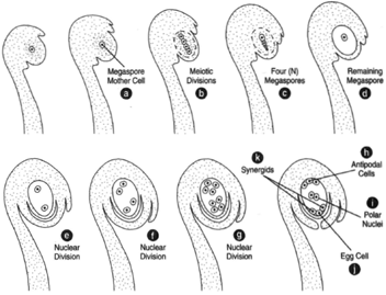 |
Figure 28-2 Female gametophyte development in an ovule. (a) Ovule with a megaspore
mother cell. (b) The first and second divisions of meiosis produce four haploid megaspores,
(c), three of which degenerate and disappear, leaving only one megaspore. (d),
(e), (f), and (g) The megaspore undergoes three nuclear divisions to produce an eightnucleated
embryo sac. (h) Three nuclei migrate to the upper end to form antipodal cells
surrounded by cell membranes. (i) Two polar nuclei remain in the center of the embryo
sac. Three nuclei migrate to the lower end (the micropylar end) and form cell membranes,
resulting in an egg cell in the center, (i), bounded by two synergids, (j). |
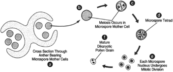 |
Figure 28-3 (a) Cross section of an anther bearing microspore mother cells. (b) and (c)
A microspore mother cell undergoes meiosis to produce a tetrad of microspores, (d). The
microspores separate (e). The nucleus of each microspore undergoes a single mitotic
division to produce a mature dikaryotic pollen grain. (g)
|
The next event is termed double fertilization. Both sperm nuclei are involved in fusions. One sperm nucleus fertilizes the egg, and the other sperm nucleus migrates to and fuses with the two polar nuclei, yielding a triploid endosperm nucleus. The zygote divides numerous times to produce the embryo sporophyte, which resides in the developing seed. At the same time, the endosperm nucleus undergoes a number of divisions to produce a reserve of nutrients. The divisions of the endosperm nucleus are not initially accompanied by cell membrane formation; the endosperm is thus coenocytic. After the endosperm has formed, cell membranes are generated, and the tissue becomes cellular. Although the events vary in different species, figure 28-6 shows the endosperm being consumed by the embryo sporophyte as the sporophyte increases in size. Whenth e seed is mature, nearly no endosperm remains.
The seed contains a 2N embryo sporophyte surrounded in early development by a 3N endosperm. The antipodal cells and synergids have degenerated. All, in turn, are surrounded by seed coats, the tissues of an older sporophyte generation. There are thus three generations of tissues present: two sporophyte generations with a gametophyte generation in between.
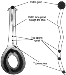 |
| Figure 28-4 At left, a male gametophyte in the pistil of a flower. At right, the male
gametophyte alone. |
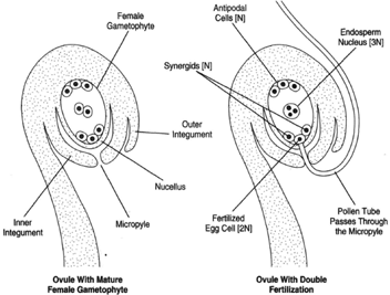 |
| Figure 28-4 At left, an ovule with a mature female gametophyte. At right, an ovule in which double fertilization has taken place. |
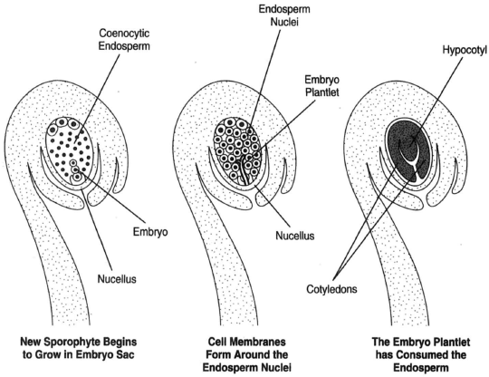 |
| Figure 28-6 At left, a new sporophyte has begun to grow in the embryo sac. At center, cell membranes have formed around the endosperm nuclei. At right, the embryo plantlet has consumed the endosperm and developed two cotyledons and the hypocotyl. |
Support our developers


