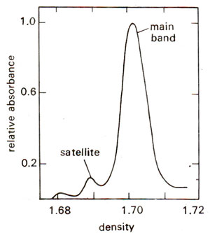
Fig. 28.9. Density centrifugation of embryonic DNA of Drosophila melanogaster, showing a main band and a satellite band.
Repetitive DNA in the form of satellite DNA
In many eukaryotic systems, highly repetitive DNA sequences of small size are found, which happen to deviate significantly from the average base composition of the total DNA. Such a fraction of DNA will have a distinct
buoyant density (measured through density centrifugation), which may be higher or lower than that of the total DNA. Therefore, this fraction can be separated as a band distinct from the main band of DNA on CsCl
2 density ultracentrifugation and is called the
satellite band (Fig. 28.9).
The satellite band is found on the left of the main band if lighter and on the right if heavier than the DNA of the main band. The DNA separated as a satellite band on ultracentrifugation is therefore, called satellite DNA and has been identified in a variety of eukaryotes including mouse, rat, guinea pig, man, fruitfly, and several dicotyledonous flowering plants. There can also be more than one satellite bands in the same organism, and in such a case they are designated as Sat-I, Sat-II, Sat-III, etc. (e.g., in
Drosophila and in humans). The proportion of DNA found in the form of satellite DNA may vary from 3-5% to as high as 40% in some cases. The lower limit is arbitrary, since less than 3% DNA can not be easily separated, without using special modified techniques.
Fig. 28.9. Density centrifugation of embryonic DNA of Drosophila melanogaster, showing a main band and a satellite band.
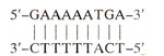
The repetitive sequences found in most of the satellite DNAs in a variety of organisms are very short and can not code for any protein. Their function is largely unknown except of ribosomal DNA found in the form of satellite DNA and constituting thousands of rRNA genes. There are many examples, where base sequences of satellite DNA have been worked out. For instance, major mouse satellite DNA consists of tandem repeats of the following sequence.
Similarly, in crab the following two satellite DNA sequence are found.
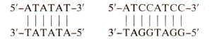
In each case, the sequences shown above are repeated millions of times. In some other cases the sequences in satellite DNA could be as long as few hundred base pairs as in humans. In such cases, sometimes, these repetitive satellite DNA sequences may not be very different from some other dispersed repetitive sequences, although in being closely clustered in tandem arrays, they differ from repetitive sequences like
Alu elements found in mammals. Two remarkable features of satellite DNA are (i) remarkable relative uniformity within the same species and (ii) great variability between otherwise closely related species. In view of these properties, it is difficult to assign any function to satellite DNA except in ribosomal RNA genes as mentioned above.
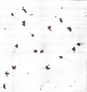
Fig. 28.10. In-situ hybridization of mouse satellite DNA showing ils location near cenlromeres.
In-situ hybridization of sat DNA
The satellite DNA often lies in heterochromatic regions of chromosomes and its location can be easily demonstrated by the technique of
in-situ or cytological hybridization. In this technique, DNA within the cell is denatured by treating the cells, that have been squashed on a cover slip. The cover slips can then be incubated in a solution of radioactively labeled satellite or any other DNA, whose location is to be determined. The sat-DNA will hybridize with denatured DNA of the cell and location can be determined by autoradiography. As shown in Figure 28.10, position of mouse sat-DNA at the chromosome ends was revealed by
in-situ hybridization. These chromosome ends also represent the centromeres in mouse.
Fig. 28.10. In-situ hybridization of mouse satellite DNA showing ils location near cenlromeres.


