Construction of a restriction map

The data of digestion by more than one endonucleases as discussed above can be utilized to arrange the sites of breakage in a defined order. This can be done by several methods, and one of them is illustrated in Figure 40.2. This method involves double digests, where we extract each fragment produced in the individual digests with either enzyme A or enzyme B and then cleave it with the other enzyme. The original DNA sample is also digested by a mixture of both the enzymes to confirm the results of individual successive digests. In Figure 40.3, we have shown the results of such an exercise. When the fragment A-2100 is retrieved and digested with enzyme B, it is cut into two fragments of 1900 bp and 200 bp. Fragment B-2500 (obtained from individual digest with enzyme B) is similarly cut into two fragments of 1900 bp and 600 bp, suggesting overlap of A-2100 and B-2500 (Fig. 40.4), in the region of the fragment of 1900bp which is obtained by one cut enzyme A and the other by enzyme B. It may be seen in Figure 40.2 that in reciprocal digests (A followed by B and B followed by A) same fragments are available. Using this information, we can find the overlapping regions in A and B. digests and find out the sites of cleavage by A and B. This will then allow us to prepare the restriction map as shown in Figure 40.5.
Technique of double digests to determine cleavage positions of DNA due to one enzyme with respect to those due to another enzyme (the two enzymes are A and B); the four gels on the extreme left show the result of electrophoresis after digesting with enzyme B each of the four fragments obtained after digestion with A ; three central gels represent results of digesting with enzyme A each of the three fragments obtained after digestion with enzyme B and the solitary gel on the right shows the results of digesting the intact DNA with both the enzymes simultaneously
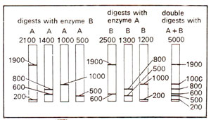
Fig. 40.2. Technique of double digests to determine cleavage positions of DNA due to one enzyme with respect to those due to another enzyme (the two enzymes are A and B); the four gels on the extreme left show the result of electrophoresis after digesting with enzyme B each of the four fragments obtained after digestion with A ; three central gels represent results of digesting with enzyme A each of the three fragments obtained after digestion with enzyme B and the solitary gel on the right shows the results of digesting the intact DNA with both the enzymes simultaneously.
The sites of cleavage by enzymes A and B in one of the four fragments, 2100 bp long obtained due to cleavage by enzyme A; the fragment has a site for enzyme B at 200 bp length from one end
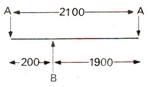
Fig. 40.3. The sites of cleavage by enzymes A and B in one of the four fragments, 2100 bp long obtained due to cleavage by enzyme A; the fragment has a site for enzyme B at 200 bp length from one end.
Reconstruction of restriction map showing restriction sites of enzymes A and B in two overlapping fragments (2100 bp fragment obtained due to enzyme A and 2500 bp fragment obtained due to B)
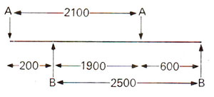
Fig. 40.4. Reconstruction of restriction map showing restriction sites of enzymes A and B in two overlapping fragments (2100 bp fragment obtained due to enzyme A and 2500 bp fragment obtained due to B).
A restriction map, in the form of linear arrangement of cleavage sites, the upper two figures show sites due to individual enzymes and the bottom figure shows arrangement of cleavage sites of both enzymes (see text for details); sites of more enzymes can be added to this map by same technique.
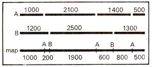
Fig. 40.5. A restriction map, in the form of linear arrangement of cleavage sites, the upper two figures show sites due to individual enzymes and the bottom figure shows arrangement of cleavage sites of both enzymes (see text for details); sites of more enzymes can be added to this map by same technique.