Nucleic Acids: Molecular Basis of Inheritance
Storage and
Transfer of Genetic
Information
Nucleic Acids: Molecular Basis of Inheritance
Cells contain two kinds of nucleic acids:
deoxyribonucleic acid (DNA), which is
the genetic material, and ribonucleic
acid (RNA), which functions in protein
synthesis. Both DNA and RNA are polymers
built of repeated units called
nucleotides. Each nucleotide contains
three parts: a sugar, a nitrogenous base, and a phosphate group. The
sugar is a pentose (5-carbon) sugar; in
DNA it is deoxyribose and in RNA it is ribose (Figure 5-11).
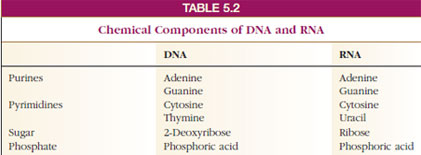
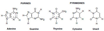
The nitrogenous bases of nucleotides are also of two types: pyrimidines, which consist of a single, 6-membered ring, and purines, which are composed of two fused rings. Both of these types of compounds contain nitrogen as well as carbon in their rings, which is why they are called “nitrogenous” bases. The purines in both RNA and DNA are adenine and guanine (Table 5-2). The pyrimidines in DNA are thymine and cytosine, and in RNA they are uracil and cytosine. The carbon atoms in the bases are numbered (for identification) according to standard biochemical notation (Figure 5-12). The carbons in the ribose and deoxyribose are also numbered, but to distinguish them from the carbons in the bases, the numbers for the carbons in the sugars are given prime signs (see Figure 5-11).
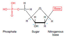
The sugar, phosphate group, and nitrogenous base are linked as shown in the generalized scheme for a nucleotide:
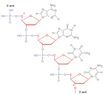
In DNA the backbone of the molecule is built of phosphoric acid and deoxyribose; to this backbone are attached the nitrogenous bases (Figure 5-13). The 5´ end of the backbone has a free phosphate group on the 5´ carbon of the ribose, and the 3´ end has a free hydroxyl group on the 3´ carbon. However, one of the most interesting and important discoveries about the nucleic acids is that DNA is not a single polynucleotide chain; rather it consists of two complementary chains that are precisely cross-linked by specific hydrogen bonding between purine and pyrimidine bases. The number of adenines is equal to the number of thymines, and the number of guanines equals the number of cytosines. This fact suggested a pairing of bases: adenine with thymine (AT) and guanine with cytosine (GC) (Figures 1-6 and 5-14).
The result is a ladder structure (Figure 5-15). The upright portions are the sugar phosphate backbones, and the connecting rungs are the paired nitrogenous bases, AT or GC. However, the ladder is twisted into a double helix with approximately 10 base pairs for each complete turn of the helix (Figure 5-16). The two DNA strands run in opposite directions, that is they are antiparallel, and the 5´ end of one strand is the 3´ end of the other. This is evident from an examination of Figure 5-16. The two strands are also complementary—the sequence of bases along one strand specifies the sequence of bases along the other strand.
The determination of the structure of DNA has been widely acclaimed as the single most important biological discovery of this century. It was based on the x-ray diffraction studies of Maurice H. F. Wilkins and Rosalind Franklin and on the ingenious proposals of Francis H. C. Crick and James D. Watson published in 1953. Watson, Crick, and Wilkins were later awarded the Nobel Prize for Physiology or Medicine for their momentous work.
RNA is similar to DNA in structure except that it consists of a single polynucleotide chain, has ribose instead of deoxyribose, and has uracil instead of thymine. The three kinds of RNA (ribosomal, transfer, and messenger) are described below.
Every time a cell divides, the structure
of DNA must be precisely copied
in the daughter cells. This is called replication (Figure 5-17). During replication,
the two strands of the double
helix unwind, and each separated
strand serves as a template against
which a complementary strand is synthesized.
That is, an enzyme (DNA
polymerase) assembles a new strand of
polynucleotides with a thymine group
going next to the adenine group in the
template strand, a guanine group next
to the cytosine group, and so on.
Nucleic Acids: Molecular Basis of Inheritance
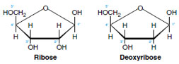 |
| Figure 5-11 Ribose and deoxyribose, the pentose sugars of nucleic acids. A carbon atom lies in each of the four corners of the pentagon (labeled 1´ to 4´). Ribose has a hydroxyl group (−OH) and a hydrogen on the number 2´ carbon; deoxyribose has two hydrogens at this position. |
The nitrogenous bases of nucleotides are also of two types: pyrimidines, which consist of a single, 6-membered ring, and purines, which are composed of two fused rings. Both of these types of compounds contain nitrogen as well as carbon in their rings, which is why they are called “nitrogenous” bases. The purines in both RNA and DNA are adenine and guanine (Table 5-2). The pyrimidines in DNA are thymine and cytosine, and in RNA they are uracil and cytosine. The carbon atoms in the bases are numbered (for identification) according to standard biochemical notation (Figure 5-12). The carbons in the ribose and deoxyribose are also numbered, but to distinguish them from the carbons in the bases, the numbers for the carbons in the sugars are given prime signs (see Figure 5-11).
The sugar, phosphate group, and nitrogenous base are linked as shown in the generalized scheme for a nucleotide:
In DNA the backbone of the molecule is built of phosphoric acid and deoxyribose; to this backbone are attached the nitrogenous bases (Figure 5-13). The 5´ end of the backbone has a free phosphate group on the 5´ carbon of the ribose, and the 3´ end has a free hydroxyl group on the 3´ carbon. However, one of the most interesting and important discoveries about the nucleic acids is that DNA is not a single polynucleotide chain; rather it consists of two complementary chains that are precisely cross-linked by specific hydrogen bonding between purine and pyrimidine bases. The number of adenines is equal to the number of thymines, and the number of guanines equals the number of cytosines. This fact suggested a pairing of bases: adenine with thymine (AT) and guanine with cytosine (GC) (Figures 1-6 and 5-14).
|
|
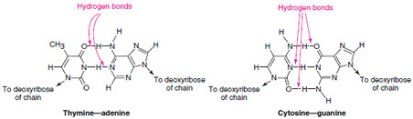 |
| Figure 5-14 Positions of hydrogen bonds between thymine and adenine and between cytosine and guanine in DNA. |
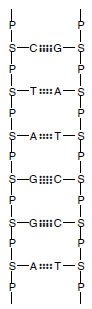
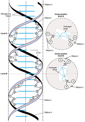
The result is a ladder structure (Figure 5-15). The upright portions are the sugar phosphate backbones, and the connecting rungs are the paired nitrogenous bases, AT or GC. However, the ladder is twisted into a double helix with approximately 10 base pairs for each complete turn of the helix (Figure 5-16). The two DNA strands run in opposite directions, that is they are antiparallel, and the 5´ end of one strand is the 3´ end of the other. This is evident from an examination of Figure 5-16. The two strands are also complementary—the sequence of bases along one strand specifies the sequence of bases along the other strand.
The determination of the structure of DNA has been widely acclaimed as the single most important biological discovery of this century. It was based on the x-ray diffraction studies of Maurice H. F. Wilkins and Rosalind Franklin and on the ingenious proposals of Francis H. C. Crick and James D. Watson published in 1953. Watson, Crick, and Wilkins were later awarded the Nobel Prize for Physiology or Medicine for their momentous work.
RNA is similar to DNA in structure except that it consists of a single polynucleotide chain, has ribose instead of deoxyribose, and has uracil instead of thymine. The three kinds of RNA (ribosomal, transfer, and messenger) are described below.
|
|
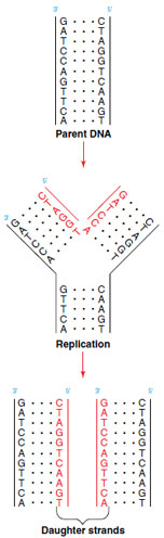 |
| Figure 5-17 Replication of DNA. The parent strands of DNA part, and DNA polymerase synthesizes daughter strands using the base sequence of parent strands as a template. The diagram shows inidirectional replication, but most DNA replication is bidirectional —proceeds in both directions at once. |
Support our developers


