Equilibrium
Equilibrium
In invertebrates, specialized sense organs for monitoring gravity and lowfrequency vibrations often appear as statocysts. Each is a simple sac lined with hair cells and containing a heavy calcareous structure, the statolith (Figure 35-28). The delicate, hairlike filaments of sensory cells are activated by the shifting position of the statolith when the animal changes position. Statocysts are found in many invertebrate phyla from radiates to arthropods. All are built on similar principles. The vertebrate organ of equilibrium is the labyrinth. It consists of two small chambers (saccule and utricle) and three semicircular canals (Figure 35-25B). The utricle and saccule are static balance organs that, like invertebrate statocysts, give information about the position of the head or body with respect to the force of gravity. As the head is tilted in one direction or another, stony accretions press on different groups of hair cells; these cells send nerve impulses to the brain, which interprets this information with reference to head position.
The semicircular canals of vertebrates are designed to respond to rotational acceleration and are relatively insensitive to linear acceleration. The three semicircular canals are at rightangles to each other, one for each axis of rotation. They are filled with fluid (endolymph), and within each canal is a bulblike enlargement, the ampulla, which contains hair cells. The hair cells are embedded in a gelatinous membrane, the cupula, which projects into the fluid. The cupula is similar in structure to the cupula of the lateral line system of fishes. When the head rotates, fluid in the canal at first tends not to move because of inertia. Since the cupula is attached, its free end is pulled in the direction opposite the direction of rotation (Figure 35-29). Bending of the cupula distorts and excites the hair cells embedded in it, and this stimulation increases the discharge rate over afferent nerve fibers leading from the ampulla to the brain. This increased discharge rate produces the sensation of rotation. Since the three canals of each ear are in different planes, acceleration in any direction stimulates at least one ampulla.
In invertebrates, specialized sense organs for monitoring gravity and lowfrequency vibrations often appear as statocysts. Each is a simple sac lined with hair cells and containing a heavy calcareous structure, the statolith (Figure 35-28). The delicate, hairlike filaments of sensory cells are activated by the shifting position of the statolith when the animal changes position. Statocysts are found in many invertebrate phyla from radiates to arthropods. All are built on similar principles. The vertebrate organ of equilibrium is the labyrinth. It consists of two small chambers (saccule and utricle) and three semicircular canals (Figure 35-25B). The utricle and saccule are static balance organs that, like invertebrate statocysts, give information about the position of the head or body with respect to the force of gravity. As the head is tilted in one direction or another, stony accretions press on different groups of hair cells; these cells send nerve impulses to the brain, which interprets this information with reference to head position.
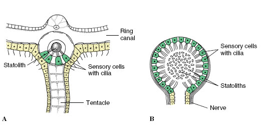 |
| Figure 35-28 Types of statocysts, static balance organs of invertebrates. A, Statocyst of the medusa of the hydrozoan Obelia. B, Statocyst of the bivalve mollusc Pecten. |
The semicircular canals of vertebrates are designed to respond to rotational acceleration and are relatively insensitive to linear acceleration. The three semicircular canals are at rightangles to each other, one for each axis of rotation. They are filled with fluid (endolymph), and within each canal is a bulblike enlargement, the ampulla, which contains hair cells. The hair cells are embedded in a gelatinous membrane, the cupula, which projects into the fluid. The cupula is similar in structure to the cupula of the lateral line system of fishes. When the head rotates, fluid in the canal at first tends not to move because of inertia. Since the cupula is attached, its free end is pulled in the direction opposite the direction of rotation (Figure 35-29). Bending of the cupula distorts and excites the hair cells embedded in it, and this stimulation increases the discharge rate over afferent nerve fibers leading from the ampulla to the brain. This increased discharge rate produces the sensation of rotation. Since the three canals of each ear are in different planes, acceleration in any direction stimulates at least one ampulla.
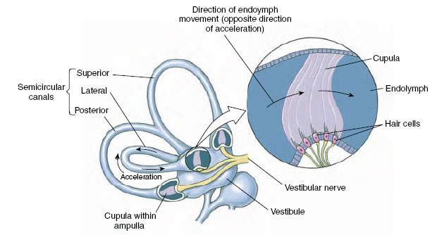 |
| Figure 35-29 How the semicircular canals respond to angular acceleration. Because of inertia, endolymph in the semicircular canal corresponding to the plane of motion moves past the cupula in a direction opposite to that of angular acceleration. Movement of the cupula stimulates hair cells. |
Support our developers


