Cellular Defenses: Phagocytosis
Cellular Defenses:
Phagocytosis
For defense against an invader an animal’s cells must recognize when a substance does not belong; they must recognize nonself. Phagocytosis illustrates nonself recognition, and it also serves as a process for removing senescent cells and cellular debris from the host. Phagocytosis occurs in almost all metazoa and is a feeding mechanism in many single-celled organisms . A cell that has this ability is a phagocyte. Phagocytes engulf a particle within an invagination of the phagocyte’s cell membrane. The invagination becomes pinched off, and the particle becomes enclosed within an intracellular vacuole. Other cytoplasmic vacuoles called lysosomes join with the particle-containing vacuole and pour in digestive enzymes to destroy the particle. Lysosomes of many phagocytes also contain enzymes that catalyze production of cytotoxic reactive oxygen intermediates (ROIs) and reactive nitrogen intermediates (RNIs). Examples of ROIs are superoxide radical (O2 −), hydrogen peroxide (H2O2), singlet oxygen (1O2), and hydroxyl radical (OH•). RNIs include nitric oxide (NO) and its oxidized forms, nitrite (NO2 −) and nitrate (NO3 −). All such intermediates are potentially toxic to invasive microorganisms or parasites.
Phagocytes and Other Defense Cells Many invertebrates have specialized cells that function as itinerant troubleshooters within the body, acting to engulf or encapsulate foreign material (see Table 37-3) and repair wounds. Such cells are variously known as amebocytes, hemocytes, or coelomocytes in different animals. If a foreign particle is small, it is engulfed by phagocytosis; but if it is larger than about 10 µm, it is usually encapsulated. Arthropods can encapsulate a foreign object by deposition of melanin around it, either from the cells of the capsule or by precipitation from the hemolymph (blood).
In vertebrates several categories of cells are capable of phagocytosis. Monocytes arise from stem cells in the bone marrow (Figure 37-1) and give rise to the mononuclear phagocyte system (reticuloendothelial [RE] system), which are phagocytic cells stationed around the body. The RE system includes macrophages in lymph nodes, spleen, and lung, Kupffer cells in sinusoids of the liver; and microglial cells in the central nervous system. Macrophages also have important roles in the specific immune response of vertebrates (see following text).
Some polymorphonuclear leukocytes (PMNs), a name that refers to the highly variable shape of their nucleus, are circulating phagocytes in blood. Another name for these leukocytes is granulocytes, which alludes to the many small granules that can be seen in their cytoplasm after treatment with appropriate stains. According to the staining properties of their granules, granulocytes are further subdivided into neutrophils, eosinophils, and basophils. Neutrophils are the most abundant, and they provide the first line of phagocytic defense in an infection. Eosinophils in normal blood account for about 2% to 5% of the total leukocytes, and basophils are the least numerous at about 0.5%. A high eosinophilia (eosinophil count in the blood) is often associated with allergic diseases and parasitic infections.
Several other kinds of cells, including basophils, are not important as phagocytes but are important cellular components of the defense system. Mast cells are basophil-like cells found in the dermis and other tissues. When they are stimulated to do so (in inflammation, basophils and mast cells release a number of pharmacologically active substances that affect surrounding cells. Lymphocytes, including T lymphocytes (T cells) and B lymphocytes (B cells), are crucial in the acquired immune response of vertebrates. Natural killer (NK) cells are lymphocytelike cells that can kill virus-infected and tumor cells in absence of antibody. They release substances onto the target-cell surface to lyse it.
For defense against an invader an animal’s cells must recognize when a substance does not belong; they must recognize nonself. Phagocytosis illustrates nonself recognition, and it also serves as a process for removing senescent cells and cellular debris from the host. Phagocytosis occurs in almost all metazoa and is a feeding mechanism in many single-celled organisms . A cell that has this ability is a phagocyte. Phagocytes engulf a particle within an invagination of the phagocyte’s cell membrane. The invagination becomes pinched off, and the particle becomes enclosed within an intracellular vacuole. Other cytoplasmic vacuoles called lysosomes join with the particle-containing vacuole and pour in digestive enzymes to destroy the particle. Lysosomes of many phagocytes also contain enzymes that catalyze production of cytotoxic reactive oxygen intermediates (ROIs) and reactive nitrogen intermediates (RNIs). Examples of ROIs are superoxide radical (O2 −), hydrogen peroxide (H2O2), singlet oxygen (1O2), and hydroxyl radical (OH•). RNIs include nitric oxide (NO) and its oxidized forms, nitrite (NO2 −) and nitrate (NO3 −). All such intermediates are potentially toxic to invasive microorganisms or parasites.
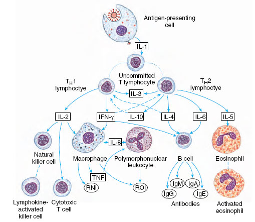 |
| Figure 37-3 Major pathways involved in cell-mediated (TH1) and humoral (TH2) immune responses as mediated by cytokines. Solid arrows indicate positive signals and broken arrows indicate inhibitory signals. Broken lines without arrows indicate path of cellular activation. IFN-γ, interferon-γ, lg, immunoglobulin; IL interleukin; TH, T helper cells; TNF, tumor necrosis factor; RNI and ROI, toxic substances released onto invader. |
Phagocytes and Other Defense Cells Many invertebrates have specialized cells that function as itinerant troubleshooters within the body, acting to engulf or encapsulate foreign material (see Table 37-3) and repair wounds. Such cells are variously known as amebocytes, hemocytes, or coelomocytes in different animals. If a foreign particle is small, it is engulfed by phagocytosis; but if it is larger than about 10 µm, it is usually encapsulated. Arthropods can encapsulate a foreign object by deposition of melanin around it, either from the cells of the capsule or by precipitation from the hemolymph (blood).
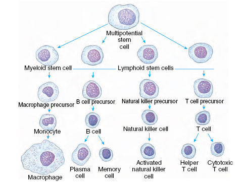 |
| Figure 37-1 Lineage of some cells active in immune response. These cells, as well as red blood cells and other white blood cells are derived from multipotential stem cells in the bone marrow. B cells mature in bone marrow and are released into blood or lymph. Precursors of T cells go through a period in the thymus gland. Precursors of macrophages circulate in blood as monocytes. |
In vertebrates several categories of cells are capable of phagocytosis. Monocytes arise from stem cells in the bone marrow (Figure 37-1) and give rise to the mononuclear phagocyte system (reticuloendothelial [RE] system), which are phagocytic cells stationed around the body. The RE system includes macrophages in lymph nodes, spleen, and lung, Kupffer cells in sinusoids of the liver; and microglial cells in the central nervous system. Macrophages also have important roles in the specific immune response of vertebrates (see following text).
Some polymorphonuclear leukocytes (PMNs), a name that refers to the highly variable shape of their nucleus, are circulating phagocytes in blood. Another name for these leukocytes is granulocytes, which alludes to the many small granules that can be seen in their cytoplasm after treatment with appropriate stains. According to the staining properties of their granules, granulocytes are further subdivided into neutrophils, eosinophils, and basophils. Neutrophils are the most abundant, and they provide the first line of phagocytic defense in an infection. Eosinophils in normal blood account for about 2% to 5% of the total leukocytes, and basophils are the least numerous at about 0.5%. A high eosinophilia (eosinophil count in the blood) is often associated with allergic diseases and parasitic infections.
Several other kinds of cells, including basophils, are not important as phagocytes but are important cellular components of the defense system. Mast cells are basophil-like cells found in the dermis and other tissues. When they are stimulated to do so (in inflammation, basophils and mast cells release a number of pharmacologically active substances that affect surrounding cells. Lymphocytes, including T lymphocytes (T cells) and B lymphocytes (B cells), are crucial in the acquired immune response of vertebrates. Natural killer (NK) cells are lymphocytelike cells that can kill virus-infected and tumor cells in absence of antibody. They release substances onto the target-cell surface to lyse it.
Support our developers


