Brain (Hindbrain, Midbrain, Forebrain)
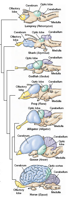 |
| Figure 35-12 Evolution of the vertebrate brain. Note the progressive increase in size of the cerebrum. The cerebellum, concerned with equilibrium and motor coordination, is largest in animals whose balance and precise motor movements are well developed (fishes, birds, and mammals). |
Unlike the spinal cord, which has changed little in structure during vertebrate evolution, the brain has changed dramatically. A primitive linear brain, as seen in fishes and amphibians, expanded to form a deeply fissured and enormously intricate brain in the lineage leading to mammals (Figure 35-12). It reaches its greatest complexity in the human brain, which contains some 35 billion nerve cells, each of which may receive information from tens of thousands of synapses at one time. The ratio between weight of the brain and that of the spinal cord affords a fair criterion of an animal’s intelligence. In fish and amphibians this ratio is approximately 1:1; in humans the ratio is 55:1—in other words, the brain is 55 times heavier than the spinal cord. Although the human brain is not the largest (the sperm whale’s brain is seven times heavier) nor the most convoluted (that of the porpoise is even more folded), it is by all odds the best in overall performance. This “great ravelled knot,” as the British physiologist Sir Charles Sherrington called the human brain, in fact may be so complex that it will never be able to understand its own function!
Although the large size of their brain undoubtedly makes humans the wisest of animals, it is apparent that they can do without much of it and still remain wise. Brain scans of persons with hydrocephalus (enlargement of the head as a result of pressure disturbances that cause the brain ventricles to enlarge many times their normal size) show that although many of them are functionally disabled, others are nearly normal. The cranium of one person with hydrocephalus was nearly filled with cerebrospinal fluid and the only remaining cerebral cortex was a thin layer of tissue,1 mm thick, pressed against the cranium.Yet this young man, with only 5% of his brain, had achieved first-class honors in mathematics at a British university and was socially normal. This and other similarly dramatic observations suggest that there is enormous redundancy and spare capacity in corticocerebral function. It also suggests that the deep structures of the brain, which are relatively spared in hydrocephalus,may peform functions once believed to be performed solely by the cortex.
Brains of early vertebrates had three principal divisions: a forebrain, or prosencephalon; a midbrain, or mesencephalon; and a hindbrain, or rhombencephalon (Figure 35-13). Each part was concerned with one or more special senses: the forebrain with smell, the midbrain with vision, and the hindbrain with hearing and balance. These primitive but very fundamental concerns of the brain have been in some instances amplified and in others reduced or overshadowed during continued evolution as sensory priorities were shaped by an animal’s habitat and way of life.
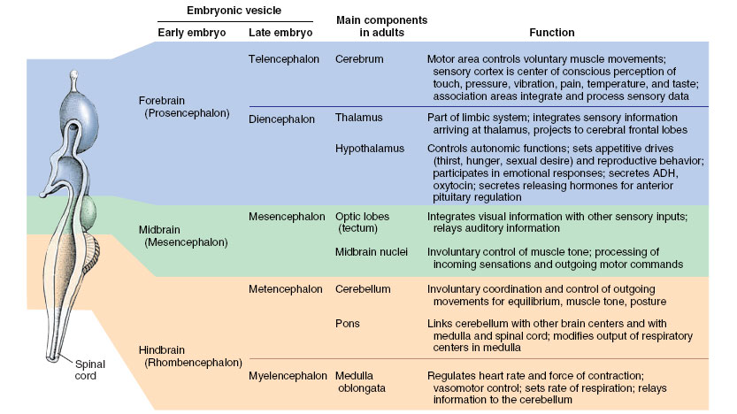 |
| Figure 35-13 Divisions of the vertebrate brain. |
Hindbrain The medulla, the most posterior division of the brain, is really a conical continuation of the spinal cord. The medulla, together with the more anterior midbrain, constitutes the “brain stem,” an area that controls numerous vital and largely subconscious activities such as heartbeat, respiration, vascular tone, gastric secretions, and swallowing. The pons, also a part of the hindbrain, contains a thick bundle of fibers that carry impulses from one side of the cerebellum to the other, and also connects both medulla and cerebellum to other brain regions.
The cerebellum, lying dorsal to the medulla, controls equilibrium, posture, and movement (Figure 35-14). Its development is directly correlated with the animal’s mode of locomotion, agility of limb movement, and balance. It is usually weakly developed in amphibians and reptiles, forms that live close to the ground, and well developed in the more agile bony fishes. It reaches its apogee in birds and mammals in which it is greatly expanded and folded. The cerebellum does not initiate movement but operates as a precision error-control center, or servomechanism, that programs a movement initiated somewhere else, such as the motor cortex of the cerebrum (Figure 35-14). Primates and especially humans, who possess a manual dexterity far surpassing that of other animals, have the most complex cerebellum. Movements of hands and fingers may involve cerebellar coordination of simultaneous contraction and relaxation of hundreds of individual muscles.
Midbrain The midbrain consists mainly of the tectum (including the optic lobes), which contains nuclei that serve as centers for visual and auditory reflexes. (In neurophysiological usage a nucleus is a small aggregation of nerve cell bodies within the central nervous system.) The midbrain has undergone little evolutionary change in structure among vertebrates but has changed markedly in function. It mediates the most complex behavior of fishes and amphibians, integrating visual, tactile, and auditory information. Such functions have been gradually assumed by the forebrain in amniotes. In mammals, the midbrain is mainly a relay center for information on its way to higher brain centers.
Forebrain Just anterior to the midbrain lie the thalamus and hypothalamus, the most posterior elements of the forebrain. The egg-shaped thalamus is a major relay station that analyzes and passes sensory information to higher brain centers. In the hypothalamus are several “housekeeping” centers that regulate body temperature, water balance, appetite, and thirst—all functions concerned with maintenance of internal constancy (homeostasis). Neurosecretory cells located in the hypothalamus produce several neurohormones (described in). The hypothalamus also contains centers for regulating reproductive function and sexual behavior, and it participates in emotional behaviors.
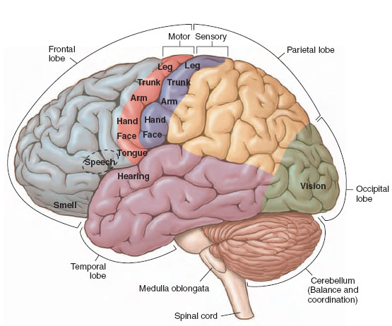 |
| Figure 35-14 External view of the human brain, showing lobes of the cerebrum and localization of major function of the cerebrum and cerebellum. |
The anterior portion of the forebrain, the cerebrum (Figure 35-14), can be divided into two anatomically distinct areas, the paleocortex and neocortex. Originally concerned with smell, it became well developed in advanced fishes and early terrestrial vertebrates, which depend on this special sense. In mammals and especially in primates the paleocortex is a deeplying area called a rhinencephalon (“nose brain”), because many of its functions depend on olfaction. Better known as the limbic system, it mediates several species-specific behaviors that relate to fulfilling needs such as feeding and sex. One region of the limbic system, the hippocampus, has been extensively studied as a site involved with spatial learning and memory. Recently, the hippocampus has gained notoriety since its neurons have been shown to divide in the adult, a previously unknown occurrence in neurons.
Although a late arrival in vertebrate evolution, the neocortex completely overshadows the paleocortex and has become so expanded that it envelops much of the forebrain and all of the midbrain (Figure 35-14). Almost all integrative activities primitively assigned to the midbrain were transferred to the neocortex, or cerebral cortex as it is usually called.
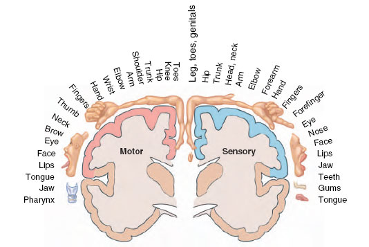 |
| Figure 35-15 Arrangement of sensory and motor cortices. Localizations of sensory terminations from different parts of the body are shown at right; origins of descending motor pathways are shown at left. The motor cortex lies in front of the sensory cortex, so the two are not superimposed. These maps grew out of the 1930s work of Canadian neurosurgeon Wilder Penfield. Recent research shows that the motor cortex is not as orderly as the map suggests; rather correspondence between cortical areas and areas of the body they control is more diffuse. |
Functions in the cerebrum have been localized by direct stimulation of exposed brains of people and experimental animals, postmortem examination of persons suffering from various lesions, and surgical removal of specific brain areas in experimental animals. The cortex contains discrete motor and sensory areas (Figures 35-14 and 35-15) as well as large “silent” regions, called association areas, concerned with memory, judgment, reasoning, and other integrative functions. These regions are not directly connected to sense organs or muscles.
Thus in mammals, and especially in humans, separate parts of the brain mediate conscious and unconscious functions. The unconscious mind, all of the brain except the cerebral cortex, governs numerous vital functions that are removed from conscious control: respiration, blood pressure, heart rate, hunger, thirst, temperature balance, salt balance, sexual drive, and basic (sometimes irrational) emotions. It is also a complex endocrine gland that regulates and receives feedback from the body’s subservient endocrine system. The conscious mind, the cerebral cortex, is the site of higher mental activities (for example, planning and reasoning), memory, and integration of sensory information. Memory appears to transcend all parts of the brain rather than being a property of any particular part of the brain as was once believed.
The right and left hemispheres of the cerebral cortex are bridged through the corpus callosum, a neural connection through which the two hemispheres are able to transfer information and coordinate mental activities. In humans, the two hemispheres of the brain are specialized for entirely different functions: the left hemisphere for language development, mathematical and learning capabilities, and sequential thought processes; and the right hemisphere for spatial, musical, artistic, intuitive, and perceptual activities. Each hemisphere also controls the opposite side of the body. It has been known for a long time that even extensive damage to the right hemisphere may cause varying degrees of leftsided paralysis but has little effect on intellect and speech. Conversely, damage to the left hemisphere usually causes loss of speech and may have disastrous effects on intellect. Since these differences in brain symmetry and function exist at birth, they appear to be inborn rather than the result of developmental or environmental effects as once believed.
Hemispheric specialization has long been considered a unique human trait, but was recently discovered in the brains of songbirds in which one side of the brain is specialized for song production.
Support our developers


