IMVIC Test
Differentiation of the principal groups of enterobacteriaceae can be accomplished on the basis of their biochemical properties and enzymeatic reactions in the presence of specific substrates. The IMVIC series of test indole, methyl red, Voges-Proskauer, and citrate utilization can be used.Indole Test
Aim
To detect the production of indole from the degradation of the amino acid tryptophan.
Introduction
Amino acids are the basic constituents of many proteins that compose living organisms. Some microorganisms degrade amino acids to yield energy in a variety of end products of ammonia, indole acid, and water. Many reactions that involve degradation of amino acids are used for classifying the enterobacteriaceae.
Certain amino acids degrading bacteria, like E. coli, have the ability to degrade the amino acids tryptophan into indole and pyruvic acid. This hydrolysis is brought about by the enzyme tryptophanase.
Principle
Indole is a nitrogen-containing compound formed from the degradation of the amino acid tryptophan. The indole test is important because only certain bacteria form indole. Indole can be easily detected with Kovac’s reagent. After the addition of the reagent and mixing the contents, the tube is allowed to stand. The alcohol layer gets separated from this aqueous layer and, upon standing, the reddening of the alcohol layer shows that Indole is present in the culture. Thus, the formation of the red layer at the top of the culture indicates the positive test. Pure tryptophan is not usually used in the test. Instead, tryptone is used as a substrate because it contains tryptophan.
Materials
- Sterilized test tubes
- Conical flasks
- Pipettes
- Glass rod
- Test culture
- Kovac’s reagent
- Tryptone broth
Procedure
- Preparation of tryptone broth:
Ingredients: Tryptone – 10 gms
Distilled water – 1000 mL
Distribute 5 mL of the broth into the test tubes and plug with cotton plugs.
Sterilize it at 121°C for 15 minutes. - Preparation of Kovac’s reagent:
N-amyl alcohol – 75 mL
Concentrated HCl – 25 mL
P-dimethylaminebenzaldehyde – 5 g. - Inoculate the tubes with the test bacterial culture.
- Incubate all the tubes for 48 hours at 37°C.
- Test for indole – Add 0.3 mL of Kovac’s reagent to each test tube. Mix well by rotating the tubes between your hands. The formation of a red layer at the top of the culture indicates a positive test.
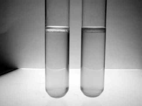 |
Figure 48 Test for Indole. |
Result
Observe your experimental result.
Discussion
One large family of Gram-negative bacteria that exhibits a considerable degree of relatedness is the enterobacteriaceae. This group is probably the most common one isolated in clinical specimens, sometimes as normal flora, sometimes as agents of diseases. The most important ones are E.coli, Klebsiella, Proteus, Enterobacter, etc.
The IMVIC test is used to identify the level of genus i.e., indole, methyl red, the Voges-Proskauer test, and the citrate test. The “I” is for pronunciation. This is a traditional panel that can be used to differentiate among several genera.
The indole test indicates the capacity of an isolate to cleave a compound indole of the amino acid tryptophan. Here, a bright ring is formed on the surface of the tube upon the addition of Kovac’s reagent if it is a positive test— or else the tube remains yellow.
Methyl Red Test
Aim
To detect the production of acids during the formation of sugars.
Introduction
Bacteria such as E.coli and Proteus, ferment glucose to produce a large amount of lactic acid, acetic acids, succinic acid, and formic acid. Carbon dioxide, hydrogen, and ethyl alcohol are also formed. When the organisms do not convert the acidic product to neutral product, the pH of the medium is 5 or less. Such acids are called mixed-acid fermenters.
Methyl red Voges-Proskauer medium is essentially a glucose broth with some buffered peptone and dipotassium phosphate. It is inoculated with test organisms. Methyl red indicator which is yellow at a pH of 5.2 and red at a pH of 4.5 is added to the culture. If the methyl red indicator turns red, then it indicates that the organism is a mixed-acid fermenter—a positive reaction. The yellow color after the addition of methyl red indicator indicates a negative reaction.
Materials
- Methyl red indicator
- Sterilized test tubes
- Conical flasks
- Glass rods
- Cotton plugs
- Test culture
- MRVP media
Procedure
- Preparation of methyl red indicator—Dissolve about 0.2 gm of methyl red in 500 mL of 95% ethyl alcohol and add 500 mL of distilled water and filter.
- Preparation of MRVP media – (glucose phosphate broth)
Dipotassium hydrogen phosphate (K2HPO4) – 5 gms
Peptone – 5 gms Glucose – 5 gms
Distilled water – 1000 mL
Suspend all the ingredients in distilled water and gently warm. Do not alter the pH. 5 mL of media that is distributed in plugged test tubes. Sterilize at 121°C for 15 minutes. - Innoculate the tubes with the test bacterial culture (except for the control tube.)
- Incubate all the tubes at 37°C for 48 hrs. 5. Add 3-4 drops of MR indicator into each tube. A distinct red color indicates the positive test; yellow color indicates a negative test.
Result
Observe your result and interpret it.
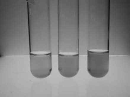 |
Figure 49 Methyl red test. |
Discussion
Studies in recent years have emphasized the complexity of the coliform group. The general practice is to classify the members. Classification is based on the results of the 4 tests—indole, MR test, VP test, and sodium citrate test.
Enterobacter cultures ferment lactose with the formation of acids, and the pH becomes less than 5. Klebsiella do not produce 2,3-butylene glycol, but others do.
E.coli, Bacillus, Proteus, and Staphylococcus a positive result as they oxidize the glucose to organic acid and stabilized the organic acid concentration. Klebsiella produces a negative result.
Voges-Proskauer Test
Aim
To detect the production of acetyl methyl carbinol (acetoin) from glucose.
Introduction
All species of Enterobacter and Serratia, as well as some species of Bacillus and Aeromonas produce a lot of 2,3-butanediol and ethyl alcohol instead of acids. These organisms are called butanediol fermenters. The production of these nonacid end products results in less lowering of the pH in methyl red Voges- Proskauer media and, thus, the test is negative.
Principles
Butanediol fermenters produces 2,3-butanediol, for which there is no satisfactory test. Acetyl methyl carbinol or acetoin (CH3CHOHCOCH3) yield 2,3-butanediol which can be easily detected with Barrit’s reagent. This test is valid, since acetoin and 2,3-butanediol are present simultaneously. In the VP test, the formation of acetyl methyl carbinol is from glucose. In the presence of alkali, atmospheric oxygen and Barrit’s reagent, small amounts of acetyl methyl carbinol present in the medium are oxidized to diacetyl, which produces a red color with guanidine residue in the media. Thus the formation of the red or pink color indicates the positive VP reaction.
Materials
- Methyl Red Voges-Proskauer medium (or glucose phosphate broth)
- Test tubes
- Pipettes
- Test cultures
- Cotton plugs
- Barrit’s reagent
Procedure
- (a) Preparation of MRVP medium or glucose phosphate broth:
Dipotassium hydrogen phosphate (K2HPO4) - 5 gms
- Peptone - 5 gms
- Glucose - 5 gms
- Distilled water - 1000 mL
Suspend all the ingredients in distilled water and gently warm. Do not alter the pH. 3 mL of the media are distributed into test tubes, which are plugged. Sterilized at 121°C.
(b) Preparation of Barrit’s reagent:
Barrit’s reagent consists of 2 solutions, i.e., solutions A and B.
Solution A is prepared by dissolving 6 gms of alpha naphthol in 100 mL of 95% ethyl alcohol.
Solution B is prepared by dissolving 16 gms of potassium hydroxide in 100 mL of water. - Inoculate the tubes with the bacterial culture.
- Incubate for 48 hours at 37°C.
- Pipette 1 mL from each culture tube into clean separate tubes. Use separate pipettes for each tubes.
- Add 18 drops (0.5 mL) of Barrit’s solution A (a-naphthol) to each tube that contains the media. Add an equal amount of solution B into the same tube.
- Shake the tubes vigorously every 30 seconds. A positive reaction is indicated by the development of a pink color, which turns red in 1–2 hours, after vigorous shaking. It is a very important step to achieve complete aeration.
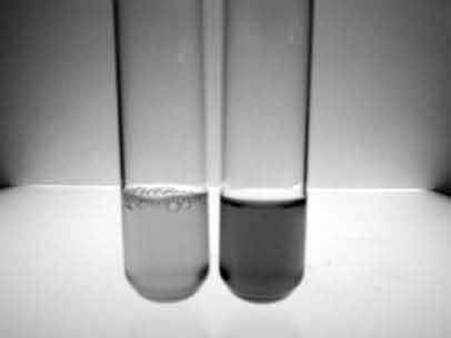 |
Figure 50 Voges–Proskauer test. |
Result
Observe your experimental result.
Discussion
Voges and Proskaver found that the addition of KOH to the cultures of organisms of the hemorrhagic septicemia by the Pasteurella group resulted in the development of a pinkish-red color, if allowed to stand for 24 hours or longer.
Harden and Walpole found that distinct differences exist in the carbohydrate metabolisms of typical enterobacters. The fermentation of glucose by the 2 organisms yielded varying amounts of products like acids, alcohol, carbon dioxide, etc. It was due to the formation of 2,3-butylene glycol and acetyl methyl carbinol by Klebsiella, but not by E.coli. Acetyl methyl carbinol in the presence of KOH and air is further oxidized to diacetyl, which in the presence of peptone produces an eosin-like color. This color is due to the guanidine nucleus of amino acids present in it.
Thus, this test is of considerable significance in testing various samples, because it disintegrates to a high degree between related enterobacters.
Citrate Utilization Test
Aim
To detect the ability to utilize citrate by microorganisms.
Introduction
Certain bacteria, such as Salmonella typhi and Escherichia aerogen, are able to use citrate as the sole source of carbon. This ability to utilize citrate as the sole source of carbon and energy is also used as a distinguishing test for certain Grods.
Principle
Simmons citrate agar, containing sodium citrate as the sole source of carbon, is used to detect if the organism can utilize citrate or not. This agar contains the indicator Bromothymol blue, which changes from green to blue when the growth of organisms causes alkalinity growth on the media. The consequent changing of color from green to blue indicates a positive test.
Materials
- Sterilized test tubes
- Conical flask
- Glass rod
- Cotton plug
- Simmons citrate agar media
Procedure
Preparation of Simmon citrate agar
- Bromothymol blue – 0.08 g
- Magnesium sulfate – 0.2 g
- Dihydrogen ammonium phosphate – 1.0 g
- Dipotassium hydrogen phosphate – 1.0 g
- Sodium citrate – 2.0 g
- Sodium chloride – 5.0 g
- Agar agar – 15 g
- Distilled water – 1000 mL
- Bromothymol blue – 40 mL
- pH – 6.8–7.0
Steps
- All the ingredients are dissolved in distilled water, heated to melt the agar, and distributed into test tubes.
- Test tubes are sterilized at 121.5°C for 15 minutes. Then the hot test tubes are placed in the slanted position.
- Test tubes are inoculated with the given bacterial culture.
- The inoculated test tubes are incubated at 37°C for 24–48 hrs.
- Growth during incubation results in the color change of the media from green to blue. This color change is regarded as a positive test
Result
Observe your experimental result.
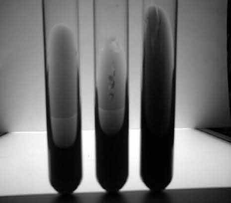 |
Figure 51 Citrate utilization test. |
Support our developers


