First Level
Peduncles are present in Colacium, an euglenophyte that forms small arborescent colonies (Figure 2.22). Its cells, with reduced flagella, are attached by their anterior pole by a peduncle consisting of an axis of neutral polysaccharides and a cortex of acid polysaccharides. Loricas are present in Trachelomonas sp. (Figure 2.23), Strombomonas verrucosa (Figure 2.24), and Ascoglena; they are very rigid, made up of mucilaginous filaments impregnated with ferric hydroxide or manganese compounds which confer an orange, brown to black coloration to the structure. These loricas fit loosely over the body proper of the cell. They possess a sharply defined collar that tapers to a more or less wide apical opening, where the flagella emerge, or possess a wide opening in one pole and attached to a substrate at the other pole, as in Ascoglena.
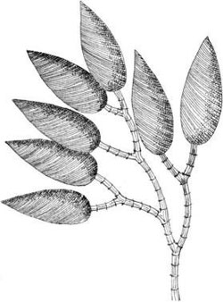
FIGURE 2.22 A small arborescent colony of Colacium sp. in which the cells are joined to one another by mucilaginous stalks.
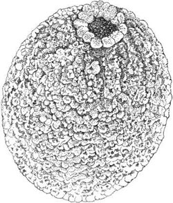
FIGURE 2.23 Lorica of Trachelomonas sp.
Beneath the mucus coating, there is the plasma membrane (Figure 2.25). This cell membrane is continuous and covers the ridges and grooves on the whole cell and can be considered the external surface of the cell. The protoplasmic face (PF) of the plasma membrane shows that the strips are covered with numerous peripheral membrane proteins of about 10 nm.
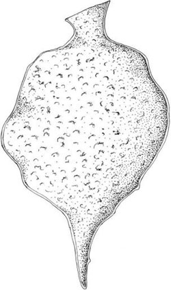
FIGURE 2.24 Lorica of Strombomonas verrucosa.
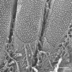
FIGURE 2.25 Deep-etching image of E. gracilis showing the mucus coating of the cell surface and the protoplasmic fracture of the cell membrane. (Bar: 0.10 µm.) (Courtesy of Dr. Pietro Lupetti.)
This peripheral cytoplasmic layer has a thickness that varies with the species. It consists of roughly twisted proteic fibers with a diameter from 10 to 15 nm arranged with an order texture or parallel striation (Figure 2.26a). The overall structure resembles the wired soul present in the tires, which gives the tire its resistance to tearing forces. Transversal fibers are detectable in some euglenoids, which connect the two longitudinal edges of the ridge of each strip (Figure 2.26b).
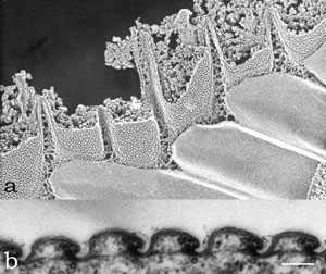
FIGURE 2.26 Deep-etching image of Euglena gracilis showing the second structural level of the pellicular complex, showing the regular texture of the internal face of the pellicle stripes (a). Transmission electron microscopy image of the pellicle of E. gracilis in transverse section showing the transversal fibers connecting the edges of successive ridges (b). (Bar: 0.10 µm)
There is a consistent number and arrangement of microtubules associated with each pellicular strip, which are continuous with those that line the flagellar canal and extend into the region of the reservoir. Within the ridge in the region of the notch there are three to five, usually four, microtubules about 25 nm diameter running parallel along each strip. Two of these are always close together and are located immediately adjacent to the notch adhering to the membrane.
The lack of protein organization in the groove regions gives higher plasticity to these zones, and together with presence of parallel microtubules in the ridge regions gives the characteristic pellicular pattern to the surface of euglenoids.
Support our developers


